4BDV | pdb_00004bdv

CRYSTAL STRUCTURE OF A TRUNCATED B-DOMAIN HUMAN FACTOR VIII


Experimental Data Snapshot

  • Method: X-RAY DIFFRACTION
  • Resolution: 3.98 Å
  • R-Value Free: 
    0.246 (Depositor), 0.220 (DCC) 
  • R-Value Work: 
    0.162 (Depositor) 
  • R-Value Observed: 
    0.166 (Depositor) 

Starting Model: experimental
View more details

wwPDB Validation 3D Report Full Report

Validation slider image for 4BDV

This is version 2.2 of the entry. See complete history

Literature

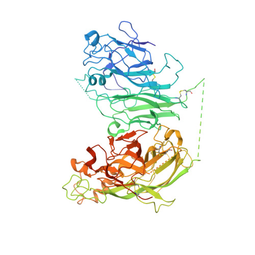
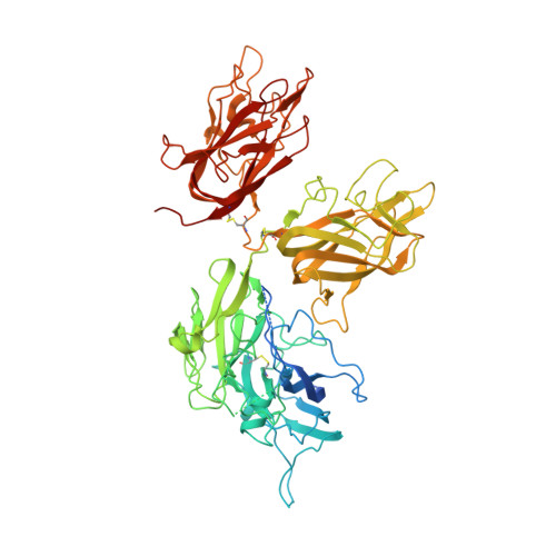
Evaluation of the Metal Binding Sites in a Recombinant Coagulation Factor Viii Identifies Two Sites with Unique Metal Binding Properties.

Svensson, L.A.Thim, L.Olsen, O.H.Nicolaisen, E.M.

(2013) Biol Chem 394: 761

  • DOI: https://doi.org/10.1515/hsz-2012-0298
  • Primary Citation Related Structures: 
    4BDV

  • PubMed Abstract: 

    Coagulation factor VIII is a glycosylated, non-covalent heterodimer consisting of a heavy chain (A1-A2-B domains) and a light chain (A3-C1-C2 domains). The association of the chains, and the stability and function of the dimer depend on the presence of metal ions. We applied X-ray fluorescence, X-ray crystallographic structure determination with anomalous signals at different wavelengths, and colorimetric measurements to evaluate the metal binding sites in a recombinant factor VIII molecule, turoctocog alfa. We identified a metal binding site in domain A3 dominated by Cu(+) binding and a site in domain A1 dominated by Zn(2+) binding.


  • Organizational Affiliation
    • Novo Nordisk A/S, Novo Nordisk Park, DK-2760 Måløv, Denmark. AnSv@novonordisk.com

Macromolecule Content 

  • Total Structure Weight: 167.98 kDa 
  • Atom Count: 9,973 
  • Modeled Residue Count: 1,218 
  • Deposited Residue Count: 1,445 
  • Unique protein chains: 2

Macromolecules

Find similar proteins by:|  3D Structure
Entity ID: 1
MoleculeChains  Sequence LengthOrganismDetailsImage
FACTOR VIIIA HEAVY CHAIN, 92 KDA ISOFORM, B DOMAIN760Homo sapiensMutation(s): 0 
UniProt & NIH Common Fund Data Resources
Find proteins for P00451 (Homo sapiens)
Explore P00451 
Go to UniProtKB:  P00451
PHAROS:  P00451
GTEx:  ENSG00000185010 
Entity Groups
Sequence Clusters30% Identity50% Identity70% Identity90% Identity95% Identity100% Identity
UniProt GroupP00451
Glycosylation
Glycosylation Sites: 1Go to GlyGen: P00451-1
Sequence Annotations
Expand
Reference Sequence
Find similar proteins by:|  3D Structure
Entity ID: 2
MoleculeChains  Sequence LengthOrganismDetailsImage
FACTOR VIIIA LIGHT CHAIN685Homo sapiensMutation(s): 0 
UniProt & NIH Common Fund Data Resources
Find proteins for P00451 (Homo sapiens)
Explore P00451 
Go to UniProtKB:  P00451
PHAROS:  P00451
GTEx:  ENSG00000185010 
Entity Groups
Sequence Clusters30% Identity50% Identity70% Identity90% Identity95% Identity100% Identity
UniProt GroupP00451
Glycosylation
Glycosylation Sites: 1Go to GlyGen: P00451-1
Sequence Annotations
Expand
Reference Sequence

Oligosaccharides

Help  
Entity ID: 3
MoleculeChains Length2D Diagram GlycosylationD Interactions
2-acetamido-2-deoxy-beta-D-glucopyranose-(1-4)-2-acetamido-2-deoxy-beta-D-glucopyranose
C
2N-Glycosylation
Glycosylation Resources
GlyTouCan: G42666HT
GlyCosmos: G42666HT
GlyGen: G42666HT
Entity ID: 4
MoleculeChains Length2D Diagram GlycosylationD Interactions
beta-D-mannopyranose-(1-3)-[beta-D-mannopyranose-(1-6)]beta-D-mannopyranose-(1-4)-2-acetamido-2-deoxy-beta-D-glucopyranose-(1-4)-2-acetamido-2-deoxy-beta-D-glucopyranose
D
5N-Glycosylation
Glycosylation Resources
GlyTouCan: G89225LT
GlyCosmos: G89225LT
GlyGen: G89225LT

Experimental Data & Validation

Experimental Data

  • Method: X-RAY DIFFRACTION
  • Resolution: 3.98 Å
  • R-Value Free:  0.246 (Depositor), 0.220 (DCC) 
  • R-Value Work:  0.162 (Depositor) 
  • R-Value Observed: 0.166 (Depositor) 
Space Group: P 41 21 2
Unit Cell:
Length ( Å )Angle ( ˚ )
a = 133.84α = 90
b = 133.84β = 90
c = 355.81γ = 90
Software Package:
Software NamePurpose
REFMACrefinement
XDSdata reduction
XSCALEdata scaling
REFMACphasing

Structure Validation

View Full Validation Report


Entry History 

Deposition Data

Revision History  (Full details and data files)

  • Version 1.0: 2013-05-15
    Type: Initial release
  • Version 1.1: 2017-01-25
    Changes: Data collection
  • Version 1.2: 2018-01-17
    Changes: Data collection
  • Version 2.0: 2020-07-29
    Type: Remediation
    Reason: Carbohydrate remediation
    Changes: Advisory, Atomic model, Data collection, Derived calculations, Other, Structure summary
  • Version 2.1: 2023-12-20
    Changes: Data collection, Database references, Refinement description, Structure summary
  • Version 2.2: 2024-10-23
    Changes: Structure summary