4BC0 | pdb_00004bc0

Structure of mouse acetylcholinesterase inhibited by CBDP (12-h soak) : Cresyl-phosphoserine adduct


Experimental Data Snapshot

  • Method: X-RAY DIFFRACTION
  • Resolution: 3.35 Å
  • R-Value Free: 
    0.208 (Depositor), 0.209 (DCC) 
  • R-Value Work: 
    0.162 (Depositor), 0.165 (DCC) 
  • R-Value Observed: 
    0.163 (Depositor) 

Starting Model: experimental
View more details

wwPDB Validation 3D Report Full Report

Validation slider image for 4BC0

Ligand Structure Quality Assessment 


This is version 1.4 of the entry. See complete history

Literature

Inhibition Pathways of the Potent Organophosphate Cbdp with Cholinesterases Revealed by X-Ray Crystallographic Snapshots and Mass Spectrometry

Carletti, E.Colletier, J.-P.Schopfer, L.M.Santoni, G.Masson, P.Lockridge, O.Nachon, F.Weik, M.

(2013) Chem Res Toxicol 26: 280

  • DOI: https://doi.org/10.1021/tx3004505
  • Primary Citation Related Structures: 
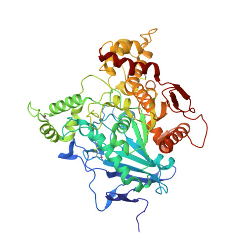
    4BBZ, 4BC0, 4BC1

  • PubMed Abstract: 

    Tri-o-cresyl-phosphate (TOCP) is a common additive in jet engine lubricants and hydraulic fluids suspected to have a role in aerotoxic syndrome in humans. TOCP is metabolized to cresyl saligenin phosphate (CBDP), a potent irreversible inhibitor of butyrylcholinesterase (BChE), a natural bioscavenger present in the bloodstream, and acetylcholinesterase (AChE), the off-switch at cholinergic synapses. Mechanistic details of cholinesterase (ChE) inhibition have, however, remained elusive. Also, the inhibition of AChE by CBDP is unexpected, from a structural standpoint, i.e., considering the narrowness of AChE active site and the bulkiness of CBDP. In the following, we report on kinetic X-ray crystallography experiments that provided 2.7-3.3 Å snapshots of the reaction of CBDP with mouse AChE and human BChE. The series of crystallographic snapshots reveals that AChE and BChE react with the opposite enantiomers and that an induced-fit rearrangement of Phe297 enlarges the active site of AChE upon CBDP binding. Mass spectrometry analysis of aging in either H(2)(16)O or H(2)(18)O furthermore allowed us to identify the inhibition steps, in which water molecules are involved, thus providing insights into the mechanistic details of inhibition. X-ray crystallography and mass spectrometry show the formation of an aged end product formed in both AChE and BChE that cannot be reactivated by current oxime-based therapeutics. Our study thus shows that only prophylactic and symptomatic treatments are viable to counter the inhibition of AChE and BChE by CBDP.


  • Organizational Affiliation
    • Institut de Biologie Structurale J.P. Ebel, Commissariat à l'Energie Atomique, 41, rue Jules Horowitz, F-38027 Grenoble, France.

Macromolecule Content 

  • Total Structure Weight: 242.89 kDa 
  • Atom Count: 17,432 
  • Modeled Residue Count: 2,161 
  • Deposited Residue Count: 2,172 
  • Unique protein chains: 1

Macromolecules

Find similar proteins by:|  3D Structure
Entity ID: 1
MoleculeChains  Sequence LengthOrganismDetailsImage
ACETYLCHOLINESTERASE
A, B, C, D
543Mus musculusMutation(s): 0 
EC: 3.1.1.7
UniProt & NIH Common Fund Data Resources
Find proteins for P21836 (Mus musculus)
Explore P21836 
Go to UniProtKB:  P21836
IMPC:  MGI:87876
Entity Groups
Sequence Clusters30% Identity50% Identity70% Identity90% Identity95% Identity100% Identity
UniProt GroupP21836
Glycosylation
Glycosylation Sites: 3Go to GlyGen: P21836-1
Sequence Annotations
Expand
Reference Sequence

Small Molecules

Ligands 4 Unique
IDChains Name / Formula / InChI Key2D Diagram3D Interactions
NAG

Query on NAG


Download:Ideal Coordinates CCD File
BA [auth D]
CA [auth D]
DA [auth D]
F [auth A]
M [auth B]
BA [auth D],
CA [auth D],
DA [auth D],
F [auth A],
M [auth B],
N [auth B],
S [auth C]
2-acetamido-2-deoxy-beta-D-glucopyranose
C8 H15 N O6
OVRNDRQMDRJTHS-FMDGEEDCSA-N
4OJ

Query on 4OJ


Download:Ideal Coordinates CCD File
AA [auth D],
E [auth A],
L [auth B],
R [auth C]
(2-methylphenyl) dihydrogen phosphate
C7 H9 O4 P
HUJOGFUFUMBXPL-UHFFFAOYSA-N
SO4

Query on SO4


Download:Ideal Coordinates CCD File
EA [auth D]
FA [auth D]
G [auth A]
GA [auth D]
H [auth A]
EA [auth D],
FA [auth D],
G [auth A],
GA [auth D],
H [auth A],
I [auth A],
J [auth A],
K [auth A],
O [auth B],
P [auth B],
Q [auth B],
V [auth C],
W [auth C]
SULFATE ION
O4 S
QAOWNCQODCNURD-UHFFFAOYSA-L
CL

Query on CL


Download:Ideal Coordinates CCD File
HA [auth D]
IA [auth D]
JA [auth D]
T [auth C]
U [auth C]
HA [auth D],
IA [auth D],
JA [auth D],
T [auth C],
U [auth C],
X [auth C],
Y [auth C],
Z [auth C]
CHLORIDE ION
Cl
VEXZGXHMUGYJMC-UHFFFAOYSA-M

Experimental Data & Validation

Experimental Data

  • Method: X-RAY DIFFRACTION
  • Resolution: 3.35 Å
  • R-Value Free:  0.208 (Depositor), 0.209 (DCC) 
  • R-Value Work:  0.162 (Depositor), 0.165 (DCC) 
  • R-Value Observed: 0.163 (Depositor) 
Space Group: P 21 21 21
Unit Cell:
Length ( Å )Angle ( ˚ )
a = 136.94α = 90
b = 174.04β = 90
c = 225.62γ = 90
Software Package:
Software NamePurpose
PHENIXrefinement
XDSdata reduction
XSCALEdata scaling
MOLREPphasing

Structure Validation

View Full Validation Report


Ligand Structure Quality Assessment 


Entry History 

Deposition Data

Revision History  (Full details and data files)

  • Version 1.0: 2013-02-06
    Type: Initial release
  • Version 1.1: 2013-02-27
    Changes: Database references
  • Version 1.2: 2020-07-29
    Type: Remediation
    Reason: Carbohydrate remediation
    Changes: Advisory, Data collection, Derived calculations, Other, Structure summary
  • Version 1.3: 2023-12-20
    Changes: Data collection, Database references, Refinement description, Structure summary
  • Version 1.4: 2024-11-20
    Changes: Structure summary