4BAL | pdb_00004bal

Thaumatin from Thaumatococcus daniellii structure in complex with the europium tris-hydroxymethyltriazoledipicolinate complex at 1.30 A resolution.


Experimental Data Snapshot

  • Method: X-RAY DIFFRACTION
  • Resolution: 1.30 Å
  • R-Value Free: 
    0.157 (Depositor), 0.155 (DCC) 
  • R-Value Work: 
    0.135 (Depositor), 0.134 (DCC) 
  • R-Value Observed: 
    0.136 (Depositor) 

Starting Model: experimental
View more details

wwPDB Validation 3D Report Full Report

Validation slider image for 4BAL

Ligand Structure Quality Assessment 


This is version 1.5 of the entry. See complete history

Literature

Clicked Europium Dipicolinate Complexes for Protein X-Ray Structure Determination.

Talon, R.Nauton, L.Canet, J.-L.Kahn, R.Girard, E.Gautier, A.

(2012) Chem Commun (Camb) 48: 11886

  • DOI: https://doi.org/10.1039/c2cc36982f
  • Primary Citation Related Structures: 
    4BAD, 4BAF, 4BAL, 4BAP, 4BAR

  • PubMed Abstract: 

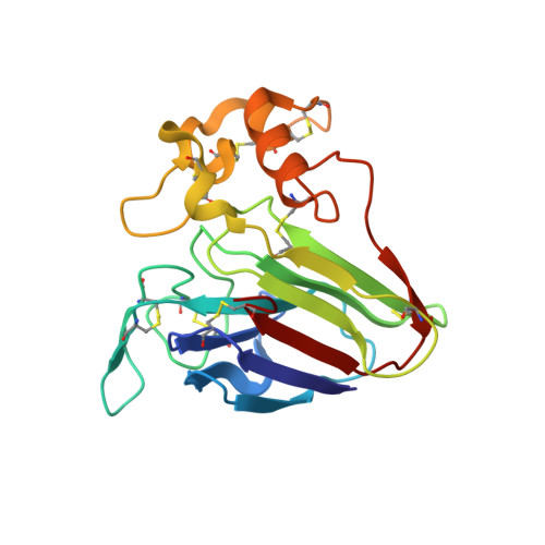
    New trisdipicolinic acid-lanthanide complexes are reported as phasing agents for X-ray crystallography of proteins. It is demonstrated that CuAAC modifications allow protein co-crystallization with low concentration of lanthanide complexes leading to an accurate structure determination.


  • Organizational Affiliation
    • CEA, DSV, Institut de Biologie Structurale (IBS), 41 rue Jules Horowitz, Grenoble F-38027, France.

Macromolecule Content 

  • Total Structure Weight: 23.17 kDa 
  • Atom Count: 2,020 
  • Modeled Residue Count: 207 
  • Deposited Residue Count: 207 
  • Unique protein chains: 1

Macromolecules

Find similar proteins by:|  3D Structure
Entity ID: 1
MoleculeChains  Sequence LengthOrganismDetailsImage
THAUMATIN-1207Thaumatococcus danielliiMutation(s): 0 
UniProt
Find proteins for P02883 (Thaumatococcus daniellii)
Explore P02883 
Go to UniProtKB:  P02883
Entity Groups
Sequence Clusters30% Identity50% Identity70% Identity90% Identity95% Identity100% Identity
UniProt GroupP02883
Sequence Annotations
Expand
Reference Sequence

Experimental Data & Validation

Experimental Data

  • Method: X-RAY DIFFRACTION
  • Resolution: 1.30 Å
  • R-Value Free:  0.157 (Depositor), 0.155 (DCC) 
  • R-Value Work:  0.135 (Depositor), 0.134 (DCC) 
  • R-Value Observed: 0.136 (Depositor) 
Space Group: P 41 21 2
Unit Cell:
Length ( Å )Angle ( ˚ )
a = 57.988α = 90
b = 57.988β = 90
c = 150.184γ = 90
Software Package:
Software NamePurpose
PHENIXrefinement
XDSdata reduction
SCALAdata scaling
PHASERphasing

Structure Validation

View Full Validation Report


Ligand Structure Quality Assessment 


Entry History 

Deposition Data

Revision History  (Full details and data files)

  • Version 1.0: 2012-11-14
    Type: Initial release
  • Version 1.1: 2012-11-28
    Changes: Database references
  • Version 1.2: 2015-04-01
    Changes: Database references
  • Version 1.3: 2019-03-06
    Changes: Data collection, Derived calculations, Experimental preparation, Other
  • Version 1.4: 2023-12-20
    Changes: Data collection, Database references, Derived calculations, Other, Refinement description
  • Version 1.5: 2024-10-16
    Changes: Structure summary