Structural Studies with Pseudomonas and Chromobacterium [Omega]-Aminotransferases Provide Insights Into Their Differing Substrate Specificity.
Sayer, C., Isupov, M.N., Westlake, A., Littlechild, J.A.(2013) Acta Crystallogr D Biol Crystallogr 69: 564
- PubMed: 23519665 Search on PubMedSearch on PubMed Central
- DOI: https://doi.org/10.1107/S0907444912051670
- Primary Citation Related Structures:
4AH3, 4B98, 4B9B, 4BA4, 4BA5 - PubMed Abstract:
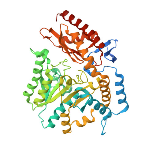
The crystal structures and inhibitor complexes of two industrially important ω-aminotransferase enzymes from Pseudomonas aeruginosa and Chromobacterium violaceum have been determined in order to understand the differences in their substrate specificity. The two enzymes share 30% sequence identity and use the same amino acceptor, pyruvate; however, the Pseudomonas enzyme shows activity towards the amino donor β-alanine, whilst the Chromobacterium enzyme does not. Both enzymes show activity towards S-α-methylbenzylamine (MBA), with the Chromobacterium enzyme having a broader substrate range. The crystal structure of the P. aeruginosa enzyme has been solved in the holo form and with the inhibitor gabaculine bound. The C. violaceum enzyme has been solved in the apo and holo forms and with gabaculine bound. The structures of the holo forms of both enzymes are quite similar. There is little conformational difference observed between the inhibitor complex and the holoenzyme for the P. aeruginosa aminotransferase. In comparison, the crystal structure of the C. violaceum gabaculine complex shows significant structural rearrangements from the structures of both the apo and holo forms of the enzyme. It appears that the different rigidity of the protein scaffold contributes to the substrate specificity observed for the two ω-aminotransferases.
- Henry Wellcome Building for Biocatalysis, Biosciences, College of Life and Environmental Sciences, University of Exeter, Exeter, England.
Organizational Affiliation: