Divergent structure-activity relationships of structurally similar acetylcholinesterase inhibitors.
Andersson, C.D., Forsgren, N., Akfur, C., Allgardsson, A., Berg, L., Engdahl, C., Qian, W., Ekstrom, F., Linusson, A.(2013) J Med Chem 56: 7615-7624
- PubMed: 23984975 Search on PubMed
- DOI: https://doi.org/10.1021/jm400990p
- Primary Citation Related Structures:
4B7Z, 4B80, 4B81, 4B82, 4B83, 4B84, 4B85, 4BTL - PubMed Abstract:
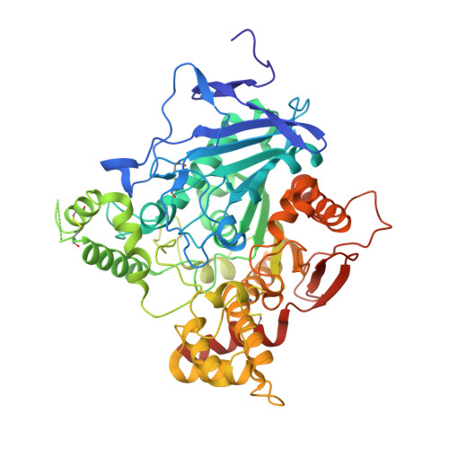
The molecular interactions between the enzyme acetylcholinesterase (AChE) and two compound classes consisting of N-[2-(diethylamino)ethyl]benzenesulfonamides and N-[2-(diethylamino)ethyl]benzenemethanesulfonamides have been investigated using organic synthesis, enzymatic assays, X-ray crystallography, and thermodynamic profiling. The inhibitors' aromatic properties were varied to establish structure-activity relationships (SAR) between the inhibitors and the peripheral anionic site (PAS) of AChE. The two structurally similar compound classes proved to have distinctly divergent SARs in terms of their inhibition capacity of AChE. Eight X-ray structures revealed that the two sets have different conformations in PAS. Furthermore, thermodynamic profiles of the binding between compounds and AChE revealed class-dependent differences of the entropy/enthalpy contributions to the free energy of binding. Further development of the entropy-favored compound class resulted in the synthesis of the most potent inhibitor and an extension beyond the established SARs. The divergent SARs will be utilized to develop reversible inhibitors of AChE into reactivators of nerve agent-inhibited AChE.
- Department of Chemistry, Umeå University , SE-901 87 Umeå, Sweden.
Organizational Affiliation: