4B3W | pdb_00004b3w

Crystal structure of human cytoglobin H(E7)Q mutant


Experimental Data Snapshot

  • Method: X-RAY DIFFRACTION
  • Resolution: 2.80 Å
  • R-Value Free: 
    0.277 (Depositor), 0.278 (DCC) 
  • R-Value Work: 
    0.207 (Depositor), 0.207 (DCC) 
  • R-Value Observed: 
    0.214 (Depositor) 

Starting Model: experimental
View more details

wwPDB Validation 3D Report Full Report

Validation slider image for 4B3W

Ligand Structure Quality Assessment 


This is version 1.1 of the entry. See complete history

Literature

Co Rebinding Kinetics and Molecular Dynamics Simulations Highlight Dynamic Regulation of Internal Cavities in Human Cytoglobin.

Gabba, M.Abbruzzetti, S.Spyrakis, F.Forti, F.Bruno, S.Mozzarelli, A.Luque, F.J.Viappiani, C.Cozzini, P.Nardini, M.Germani, F.Bolognesi, M.Moens, L.Dewilde, S.

(2013) PLoS One 8: 49770

  • DOI: https://doi.org/10.1371/journal.pone.0049770
  • Primary Citation Related Structures: 
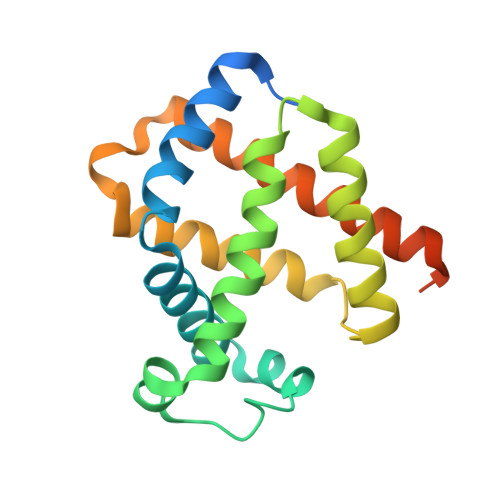
    4B3W

  • PubMed Abstract: 

    Cytoglobin (Cygb) was recently discovered in the human genome and localized in different tissues. It was suggested to play tissue-specific protective roles, spanning from scavenging of reactive oxygen species in neurons to supplying oxygen to enzymes in fibroblasts. To shed light on the functioning of such versatile machinery, we have studied the processes supporting transport of gaseous heme ligands in Cygb. Carbon monoxide rebinding shows a complex kinetic pattern with several distinct reaction intermediates, reflecting rebinding from temporary docking sites, second order recombination, and formation (and dissociation) of a bis-histidyl heme hexacoordinated reaction intermediate. Ligand exit to the solvent occurs through distinct pathways, some of which exploit temporary docking sites. The remarkable change in energetic barriers, linked to heme bis-histidyl hexacoordination by HisE7, may be responsible for active regulation of the flux of reactants and products to and from the reaction site on the distal side of the heme. A substantial change in both protein dynamics and inner cavities is observed upon transition from the CO-liganded to the pentacoordinated and bis-histidyl hexacoordinated species, which could be exploited as a signalling state. These findings are consistent with the expected versatility of the molecular activity of this protein.


  • Organizational Affiliation
    • Institute of Complex Systems - Molekulare Biophysik (ICS-5) Forschungszentrum Jülich, Jülich, Germany.

Macromolecule Content 

  • Total Structure Weight: 44.55 kDa 
  • Atom Count: 2,610 
  • Modeled Residue Count: 308 
  • Deposited Residue Count: 380 
  • Unique protein chains: 1

Macromolecules

Find similar proteins by:|  3D Structure
Entity ID: 1
MoleculeChains  Sequence LengthOrganismDetailsImage
CYTOGLOBIN
A, B
190Homo sapiensMutation(s): 3 
EC: 1.11.1 (UniProt), 1.14.12 (UniProt), 1.15.1.1 (UniProt), 1.7 (UniProt)
UniProt & NIH Common Fund Data Resources
Find proteins for Q8WWM9 (Homo sapiens)
Explore Q8WWM9 
Go to UniProtKB:  Q8WWM9
PHAROS:  Q8WWM9
GTEx:  ENSG00000161544 
Entity Groups
Sequence Clusters30% Identity50% Identity70% Identity90% Identity95% Identity100% Identity
UniProt GroupQ8WWM9
Sequence Annotations
Expand
Reference Sequence

Small Molecules

Ligands 4 Unique
IDChains Name / Formula / InChI Key2D Diagram3D Interactions
HEM

Query on HEM


Download:Ideal Coordinates CCD File
C [auth A],
F [auth B]
PROTOPORPHYRIN IX CONTAINING FE
C34 H32 Fe N4 O4
KABFMIBPWCXCRK-RGGAHWMASA-L
FC6

Query on FC6


Download:Ideal Coordinates CCD File
E [auth A],
H [auth B]
HEXACYANOFERRATE(3-)
C6 Fe N6
HCMVSLMENOCDCK-UHFFFAOYSA-N
ACT

Query on ACT


Download:Ideal Coordinates CCD File
I [auth B]ACETATE ION
C2 H3 O2
QTBSBXVTEAMEQO-UHFFFAOYSA-M
CYN

Query on CYN


Download:Ideal Coordinates CCD File
D [auth A],
G [auth B]
CYANIDE ION
C N
XFXPMWWXUTWYJX-UHFFFAOYSA-N

Experimental Data & Validation

Experimental Data

  • Method: X-RAY DIFFRACTION
  • Resolution: 2.80 Å
  • R-Value Free:  0.277 (Depositor), 0.278 (DCC) 
  • R-Value Work:  0.207 (Depositor), 0.207 (DCC) 
  • R-Value Observed: 0.214 (Depositor) 
Space Group: P 21 21 21
Unit Cell:
Length ( Å )Angle ( ˚ )
a = 48.844α = 90
b = 70.054β = 90
c = 102.072γ = 90
Software Package:
Software NamePurpose
REFMACrefinement
MOSFLMdata reduction
SCALAdata scaling
PHASERphasing

Structure Validation

View Full Validation Report


Ligand Structure Quality Assessment 


Entry History 

Revision History  (Full details and data files)

  • Version 1.0: 2013-01-23
    Type: Initial release
  • Version 1.1: 2023-12-20
    Changes: Data collection, Database references, Derived calculations, Other, Refinement description