4B3F

crystal structure of Ighmbp2 helicase


Experimental Data Snapshot

  • Method: X-RAY DIFFRACTION
  • Resolution: 2.50 Å
  • R-Value Free: 0.252 
  • R-Value Work: 0.190 
  • R-Value Observed: 0.193 

wwPDB Validation   3D Report Full Report


This is version 1.3 of the entry. See complete history


Literature

The Ighmbp2 Helicase Structure Reveals the Molecular Basis for Disease-Causing Mutations in Dmsa1.

Lim, S.C.Bowler, M.W.Lai, T.F.Song, H.

(2012) Nucleic Acids Res 40: 11009

  • DOI: https://doi.org/10.1093/nar/gks792
  • Primary Citation of Related Structures:  
    4B3F, 4B3G

  • PubMed Abstract: 

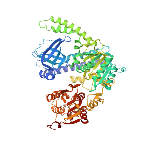
    Mutations in immunoglobulin µ-binding protein 2 (Ighmbp2) cause distal spinal muscular atrophy type 1 (DSMA1), an autosomal recessive disease that is clinically characterized by distal limb weakness and respiratory distress. However, despite extensive studies, the mechanism of disease-causing mutations remains elusive. Here we report the crystal structures of the Ighmbp2 helicase core with and without bound RNA. The structures show that the overall fold of Ighmbp2 is very similar to that of Upf1, a key helicase involved in nonsense-mediated mRNA decay. Similar to Upf1, domains 1B and 1C of Ighmbp2 undergo large conformational changes in response to RNA binding, rotating 30° and 10°, respectively. The RNA binding and ATPase activities of Ighmbp2 are further enhanced by the R3H domain, located just downstream of the helicase core. Mapping of the pathogenic mutations of DSMA1 onto the helicase core structure provides a molecular basis for understanding the disease-causing consequences of Ighmbp2 mutations.


  • Organizational Affiliation

    Institute of Molecular and Cell Biology, Proteos, Singapore 138673, Singapore.


Macromolecules
Find similar proteins by:  (by identity cutoff)  |  3D Structure
Entity ID: 1
MoleculeChains Sequence LengthOrganismDetailsImage
DNA-BINDING PROTEIN SMUBP-2A [auth X]646Homo sapiensMutation(s): 0 
EC: 3.6.4.12 (PDB Primary Data), 3.6.4.13 (PDB Primary Data)
UniProt & NIH Common Fund Data Resources
Find proteins for P38935 (Homo sapiens)
Explore P38935 
Go to UniProtKB:  P38935
PHAROS:  P38935
GTEx:  ENSG00000132740 
Entity Groups  
Sequence Clusters30% Identity50% Identity70% Identity90% Identity95% Identity100% Identity
UniProt GroupP38935
Sequence Annotations
Expand
  • Reference Sequence
Small Molecules
Ligands 1 Unique
IDChains Name / Formula / InChI Key2D Diagram3D Interactions
PO4
Query on PO4

Download Ideal Coordinates CCD File 
B [auth X]PHOSPHATE ION
O4 P
NBIIXXVUZAFLBC-UHFFFAOYSA-K
Experimental Data & Validation

Experimental Data

  • Method: X-RAY DIFFRACTION
  • Resolution: 2.50 Å
  • R-Value Free: 0.252 
  • R-Value Work: 0.190 
  • R-Value Observed: 0.193 
  • Space Group: C 1 2 1
Unit Cell:
Length ( Å )Angle ( ˚ )
a = 116.566α = 90
b = 76.717β = 107.33
c = 88.536γ = 90
Software Package:
Software NamePurpose
PHENIXrefinement
MOSFLMdata reduction
SCALAdata scaling
PHENIXphasing

Structure Validation

View Full Validation Report


Entry History 

Deposition Data

Revision History  (Full details and data files)

  • Version 1.0: 2012-09-26
    Type: Initial release
  • Version 1.1: 2012-12-19
    Changes: Database references
  • Version 1.2: 2018-05-02
    Changes: Data collection
  • Version 1.3: 2018-05-16
    Changes: Data collection