4B1H

Structure of human PARG catalytic domain in complex with ADP-ribose


Experimental Data Snapshot

  • Method: X-RAY DIFFRACTION
  • Resolution: 2.00 Å
  • R-Value Free: 0.241 
  • R-Value Work: 0.204 
  • R-Value Observed: 0.205 

wwPDB Validation   3D Report Full Report


Ligand Structure Quality Assessment 


This is version 1.3 of the entry. See complete history


Literature

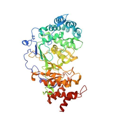
Structures of the Human Poly (Adp-Ribose) Glycohydrolase Catalytic Domain Confirm Catalytic Mechanism and Explain Inhibition by Adp-Hpd Derivatives.

Tucker, J.A.Bennett, N.Brassington, C.Durant, S.T.Hassall, G.Holdgate, G.Mcalister, M.Nissink, J.W.M.Truman, C.Watson, M.

(2012) PLoS One 7: 50889

  • DOI: https://doi.org/10.1371/journal.pone.0050889
  • Primary Citation of Related Structures:  
    4A0D, 4B1G, 4B1H, 4B1I, 4B1J

  • PubMed Abstract: 

    Poly(ADP-ribose) glycohydrolase (PARG) is the only enzyme known to catalyse hydrolysis of the O-glycosidic linkages of ADP-ribose polymers, thereby reversing the effects of poly(ADP-ribose) polymerases. PARG deficiency leads to cell death whilst PARG depletion causes sensitisation to certain DNA damaging agents, implicating PARG as a potential therapeutic target in several disease areas. Efforts to develop small molecule inhibitors of PARG activity have until recently been hampered by a lack of structural information on PARG. We have used a combination of bio-informatic and experimental approaches to engineer a crystallisable, catalytically active fragment of human PARG (hPARG). Here, we present high-resolution structures of the catalytic domain of hPARG in unliganded form and in complex with three inhibitors: ADP-ribose (ADPR), adenosine 5'-diphosphate (hydroxymethyl)pyrrolidinediol (ADP-HPD) and 8-n-octyl-amino-ADP-HPD. Our structures confirm conservation of overall fold amongst mammalian PARG glycohydrolase domains, whilst revealing additional flexible regions in the catalytic site. These new structures rationalise a body of published mutational data and the reported structure-activity relationship for ADP-HPD based PARG inhibitors. In addition, we have developed and used biochemical, isothermal titration calorimetry and surface plasmon resonance assays to characterise the binding of inhibitors to our PARG protein, thus providing a starting point for the design of new inhibitors.


  • Organizational Affiliation

    Innovative Medicines, AstraZeneca UK Ltd, Macclesfield, Cheshire, United Kingdom. julie.tucker@astrazeneca.com


Macromolecules
Find similar proteins by:  (by identity cutoff)  |  3D Structure
Entity ID: 1
MoleculeChains Sequence LengthOrganismDetailsImage
POLY(ADP-RIBOSE) GLYCOHYDROLASE531Homo sapiensMutation(s): 6 
EC: 3.2.1.143
UniProt & NIH Common Fund Data Resources
Find proteins for Q86W56 (Homo sapiens)
Explore Q86W56 
Go to UniProtKB:  Q86W56
PHAROS:  Q86W56
GTEx:  ENSG00000227345 
Entity Groups  
Sequence Clusters30% Identity50% Identity70% Identity90% Identity95% Identity100% Identity
UniProt GroupQ86W56
Sequence Annotations
Expand
  • Reference Sequence
Small Molecules
Ligands 5 Unique
IDChains Name / Formula / InChI Key2D Diagram3D Interactions
AR6
Query on AR6

Download Ideal Coordinates CCD File 
F [auth A],
G [auth A]
[(2R,3S,4R,5R)-5-(6-AMINOPURIN-9-YL)-3,4-DIHYDROXY-OXOLAN-2-YL]METHYL [HYDROXY-[[(2R,3S,4R,5S)-3,4,5-TRIHYDROXYOXOLAN-2-YL]METHOXY]PHOSPHORYL] HYDROGEN PHOSPHATE
C15 H23 N5 O14 P2
SRNWOUGRCWSEMX-ZQSHOCFMSA-N
DTV
Query on DTV

Download Ideal Coordinates CCD File 
C [auth A](2S,3S)-1,4-DIMERCAPTOBUTANE-2,3-DIOL
C4 H10 O2 S2
VHJLVAABSRFDPM-QWWZWVQMSA-N
SO4
Query on SO4

Download Ideal Coordinates CCD File 
H [auth A],
I [auth A]
SULFATE ION
O4 S
QAOWNCQODCNURD-UHFFFAOYSA-L
GOL
Query on GOL

Download Ideal Coordinates CCD File 
D [auth A],
E [auth A]
GLYCEROL
C3 H8 O3
PEDCQBHIVMGVHV-UHFFFAOYSA-N
BME
Query on BME

Download Ideal Coordinates CCD File 
B [auth A]BETA-MERCAPTOETHANOL
C2 H6 O S
DGVVWUTYPXICAM-UHFFFAOYSA-N
Modified Residues  1 Unique
IDChains TypeFormula2D DiagramParent
CSO
Query on CSO
A
L-PEPTIDE LINKINGC3 H7 N O3 SCYS
Experimental Data & Validation

Experimental Data

  • Method: X-RAY DIFFRACTION
  • Resolution: 2.00 Å
  • R-Value Free: 0.241 
  • R-Value Work: 0.204 
  • R-Value Observed: 0.205 
  • Space Group: P 21 21 21
Unit Cell:
Length ( Å )Angle ( ˚ )
a = 66.929α = 90
b = 90.656β = 90
c = 94.975γ = 90
Software Package:
Software NamePurpose
REFMACrefinement
d*TREKdata reduction
d*TREKdata scaling
AMoREphasing

Structure Validation

View Full Validation Report


Ligand Structure Quality Assessment 


Entry History 

Deposition Data

Revision History  (Full details and data files)

  • Version 1.0: 2012-12-19
    Type: Initial release
  • Version 1.1: 2013-01-16
    Changes: Database references
  • Version 1.2: 2018-04-04
    Changes: Data collection
  • Version 1.3: 2023-12-20
    Changes: Data collection, Database references, Derived calculations, Other, Refinement description, Structure summary