4AZ3 | pdb_00004az3

crystal structure of cathepsin a, complexed with 15a


Experimental Data Snapshot

  • Method: X-RAY DIFFRACTION
  • Resolution: 2.04 Å
  • R-Value Free: 
    0.215 (Depositor), 0.213 (DCC) 
  • R-Value Work: 
    0.188 (Depositor) 

Starting Model: experimental
View more details

wwPDB Validation 3D Report Full Report

Validation slider image for 4AZ3

Ligand Structure Quality Assessment 


This is version 2.2 of the entry. See complete history

Literature

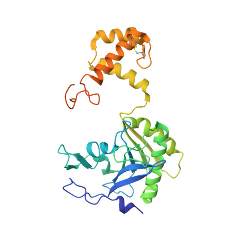
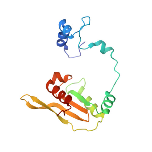
Novel Beta-Amino Acid Derivatives as Inhibitors of Cathepsin A.

Ruf, S.Buning, C.Schreuder, H.Horstick, G.Linz, W.Olpp, T.Pernerstorfer, J.Hiss, K.Kroll, K.Kannt, A.Kohlmann, M.Linz, D.Hubschle, T.Rutten, H.Wirth, K.Schmidt, T.Sadowski, T.

(2012) J Med Chem 55: 7636

  • DOI: https://doi.org/10.1021/jm300663n
  • Primary Citation Related Structures: 
    4AZ0, 4AZ3

  • PubMed Abstract: 

    Cathepsin A (CatA) is a serine carboxypeptidase distributed between lysosomes, cell membrane, and extracellular space. Several peptide hormones including bradykinin and angiotensin I have been described as substrates. Therefore, the inhibition of CatA has the potential for beneficial effects in cardiovascular diseases. Pharmacological inhibition of CatA by the natural product ebelactone B increased renal bradykinin levels and prevented the development of salt-induced hypertension. However, so far no small molecule inhibitors of CatA with oral bioavailability have been described to allow further pharmacological profiling. In our work we identified novel β-amino acid derivatives as inhibitors of CatA after a HTS analysis based on a project adapted fragment approach. The new inhibitors showed beneficial ADME and pharmacokinetic profiles, and their binding modes were established by X-ray crystallography. Further investigations led to the identification of a hitherto unknown pathophysiological role of CatA in cardiac hypertrophy. One of our inhibitors is currently undergoing phase I clinical trials.


  • Organizational Affiliation
    • Sanofi-Aventis Deutschland GmbH, Industriepark Höchst, 65926 Frankfurt, Germany. sven.ruf@sanofi.com

Macromolecule Content 

  • Total Structure Weight: 53.63 kDa 
  • Atom Count: 3,854 
  • Modeled Residue Count: 414 
  • Deposited Residue Count: 455 
  • Unique protein chains: 2

Macromolecules

Find similar proteins by:|  3D Structure
Entity ID: 1
MoleculeChains  Sequence LengthOrganismDetailsImage
LYSOSOMAL PROTECTIVE PROTEIN 32 KDA CHAIN300Homo sapiensMutation(s): 0 
EC: 3.4.16.5
UniProt & NIH Common Fund Data Resources
Find proteins for P10619 (Homo sapiens)
Explore P10619 
Go to UniProtKB:  P10619
PHAROS:  P10619
GTEx:  ENSG00000064601 
Entity Groups
Sequence Clusters30% Identity50% Identity70% Identity90% Identity95% Identity100% Identity
UniProt GroupP10619
Glycosylation
Glycosylation Sites: 1Go to GlyGen: P10619-1
Sequence Annotations
Expand
Reference Sequence
Find similar proteins by:|  3D Structure
Entity ID: 2
MoleculeChains  Sequence LengthOrganismDetailsImage
LYSOSOMAL PROTECTIVE PROTEIN 20 KDA CHAIN155Homo sapiensMutation(s): 0 
EC: 3.4.16.5
UniProt & NIH Common Fund Data Resources
Find proteins for P10619 (Homo sapiens)
Explore P10619 
Go to UniProtKB:  P10619
PHAROS:  P10619
GTEx:  ENSG00000064601 
Entity Groups
Sequence Clusters30% Identity50% Identity70% Identity90% Identity95% Identity100% Identity
UniProt GroupP10619
Glycosylation
Glycosylation Sites: 1Go to GlyGen: P10619-1
Sequence Annotations
Expand
Reference Sequence

Oligosaccharides

Help  
Entity ID: 3
MoleculeChains Length2D Diagram GlycosylationD Interactions
2-acetamido-2-deoxy-beta-D-glucopyranose-(1-4)-2-acetamido-2-deoxy-beta-D-glucopyranose
C, D
2N-Glycosylation
Glycosylation Resources
GlyTouCan: G42666HT
GlyCosmos: G42666HT
GlyGen: G42666HT

Experimental Data & Validation

Experimental Data

  • Method: X-RAY DIFFRACTION
  • Resolution: 2.04 Å
  • R-Value Free:  0.215 (Depositor), 0.213 (DCC) 
  • R-Value Work:  0.188 (Depositor) 
Space Group: C 1 2 1
Unit Cell:
Length ( Å )Angle ( ˚ )
a = 90.59α = 90
b = 102.8β = 101.87
c = 48.39γ = 90
Software Package:
Software NamePurpose
BUSTERrefinement
XDSdata reduction
XSCALEdata scaling

Structure Validation

View Full Validation Report


Ligand Structure Quality Assessment 


Entry History 

Revision History  (Full details and data files)

  • Version 1.0: 2012-09-26
    Type: Initial release
  • Version 1.1: 2019-04-03
    Changes: Data collection, Derived calculations, Experimental preparation, Other
  • Version 1.2: 2019-05-08
    Changes: Data collection, Experimental preparation
  • Version 2.0: 2020-07-29
    Type: Remediation
    Reason: Carbohydrate remediation
    Changes: Atomic model, Data collection, Derived calculations, Other, Structure summary
  • Version 2.1: 2023-12-20
    Changes: Data collection, Database references, Refinement description, Structure summary
  • Version 2.2: 2024-11-13
    Changes: Structure summary