4AYG | pdb_00004ayg

Lactobacillus reuteri N-terminally truncated glucansucrase GTF180 in orthorhombic apo-form


Experimental Data Snapshot

  • Method: X-RAY DIFFRACTION
  • Resolution: 2.00 Å
  • R-Value Free: 
    0.183 (Depositor), 0.184 (DCC) 
  • R-Value Work: 
    0.155 (Depositor), 0.156 (DCC) 
  • R-Value Observed: 
    0.156 (Depositor) 

Starting Model: experimental
View more details

wwPDB Validation 3D Report Full Report

Validation slider image for 4AYG

This is version 1.4 of the entry. See complete history

Literature

Flexibility of Truncated and Full-Length Glucansucrase Gtf180 Enzymes from Lactobacillus Reuteri 180.

Pijning, T.Vujicic-Zagar, A.Kralj, S.Dijkhuizen, L.Dijkstra, B.W.

(2014) FEBS J 281: 2159

  • DOI: https://doi.org/10.1111/febs.12769
  • Primary Citation Related Structures: 
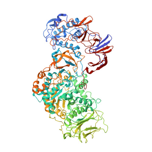
    4AYG

  • PubMed Abstract: 

    Glucansucrase enzymes synthesize high-molecular-mass extracellular α-glucan polysaccharides from sucrose. Previously, the crystal structure of truncated glucansucrase glucosyltransferase (GTF)180-ΔN from Lactobacillus reuteri 180 (lacking the N-terminal domain) revealed an elongated overall structure with two remote domains (IV and V) extending away from the core. By contrast, a new crystal form of the α-1,6/α-1,3 specific glucansucrase GTF180-ΔN shows an approximate 120(o) rotation of domain V about a hinge located between domains IV and V, giving a much more compact structure than before. Positional variability of domain V in solution is confirmed by small angle X-ray scattering experiments and rigid-body ensemble calculations. In addition, small angle X-ray scattering measurements of full-length GTF180 also provide the first structural data for a full-length glucansucrase, showing that the enzyme has an almost symmetric boomerang-like molecular shape, with a bend likely located between domains IV and V. The ~ 700-residue N-terminal domain, which is not present in the crystal structures, extends away from domain V and the catalytic core of the enzyme. We conclude that, as a result of the hinge region, in solution, GTF180-ΔN (and likely also the full-length GTF180) shows conformational flexibility; this may be a general feature of GH70 glucansucrases. • Structural data for GTF180-ΔN II have been deposited in the Protein Data Bank under accession code 4AYG.


  • Organizational Affiliation
    • Laboratory of Biophysical Chemistry, Groningen Biomolecular Sciences and Biotechnology Institute (GBB), University of Groningen, The Netherlands.

Macromolecule Content 

  • Total Structure Weight: 241.53 kDa 
  • Atom Count: 19,034 
  • Modeled Residue Count: 2,055 
  • Deposited Residue Count: 2,078 
  • Unique protein chains: 1

Macromolecules

Find similar proteins by:|  3D Structure
Entity ID: 1
MoleculeChains  Sequence LengthOrganismDetailsImage
GLUCANSUCRASE
A, B
1,039Limosilactobacillus reuteriMutation(s): 0 
EC: 2.4.1.5
UniProt
Find proteins for Q5SBN3 (Limosilactobacillus reuteri)
Explore Q5SBN3 
Go to UniProtKB:  Q5SBN3
Entity Groups
Sequence Clusters30% Identity50% Identity70% Identity90% Identity95% Identity100% Identity
UniProt GroupQ5SBN3
Sequence Annotations
Expand
Reference Sequence

Small Molecules

Ligands 5 Unique
IDChains Name / Formula / InChI Key2D Diagram3D Interactions
SO4

Query on SO4


Download:Ideal Coordinates CCD File
BC [auth B]
CC [auth B]
DB [auth A]
DC [auth B]
EC [auth B]
BC [auth B],
CC [auth B],
DB [auth A],
DC [auth B],
EC [auth B],
FC [auth B],
GC [auth B],
HC [auth B],
IC [auth B],
JC [auth B],
KA [auth A],
KC [auth B],
LA [auth A],
MA [auth A],
NA [auth A],
OA [auth A],
PA [auth A],
QA [auth A],
RA [auth A],
SA [auth A],
TA [auth A]
SULFATE ION
O4 S
QAOWNCQODCNURD-UHFFFAOYSA-L
GOL

Query on GOL


Download:Ideal Coordinates CCD File
AA [auth A]
AC [auth B]
BA [auth A]
CA [auth A]
D [auth A]
AA [auth A],
AC [auth B],
BA [auth A],
CA [auth A],
D [auth A],
DA [auth A],
E [auth A],
EA [auth A],
F [auth A],
FA [auth A],
FB [auth B],
G [auth A],
GA [auth A],
GB [auth B],
H [auth A],
HA [auth A],
HB [auth B],
I [auth A],
IA [auth A],
IB [auth B],
J [auth A],
JA [auth A],
JB [auth B],
K [auth A],
KB [auth B],
L [auth A],
LB [auth B],
M [auth A],
MB [auth B],
N [auth A],
NB [auth B],
O [auth A],
OB [auth B],
P [auth A],
PB [auth B],
Q [auth A],
QB [auth B],
R [auth A],
RB [auth B],
S [auth A],
SB [auth B],
T [auth A],
TB [auth B],
U [auth A],
UB [auth B],
V [auth A],
VB [auth B],
W [auth A],
WB [auth B],
X [auth A],
XB [auth B],
Y [auth A],
YB [auth B],
Z [auth A],
ZB [auth B]
GLYCEROL
C3 H8 O3
PEDCQBHIVMGVHV-UHFFFAOYSA-N
IPA

Query on IPA


Download:Ideal Coordinates CCD File
CB [auth A],
QC [auth B]
ISOPROPYL ALCOHOL
C3 H8 O
KFZMGEQAYNKOFK-UHFFFAOYSA-N
ACY

Query on ACY


Download:Ideal Coordinates CCD File
AB [auth A]
BB [auth A]
LC [auth B]
MC [auth B]
NC [auth B]
AB [auth A],
BB [auth A],
LC [auth B],
MC [auth B],
NC [auth B],
OC [auth B],
PC [auth B],
UA [auth A],
VA [auth A],
WA [auth A],
XA [auth A],
YA [auth A],
ZA [auth A]
ACETIC ACID
C2 H4 O2
QTBSBXVTEAMEQO-UHFFFAOYSA-N
CA

Query on CA


Download:Ideal Coordinates CCD File
C [auth A],
EB [auth B]
CALCIUM ION
Ca
BHPQYMZQTOCNFJ-UHFFFAOYSA-N

Experimental Data & Validation

Experimental Data

  • Method: X-RAY DIFFRACTION
  • Resolution: 2.00 Å
  • R-Value Free:  0.183 (Depositor), 0.184 (DCC) 
  • R-Value Work:  0.155 (Depositor), 0.156 (DCC) 
  • R-Value Observed: 0.156 (Depositor) 
Space Group: P 21 21 21
Unit Cell:
Length ( Å )Angle ( ˚ )
a = 92.792α = 90
b = 147.363β = 90
c = 244.374γ = 90
Software Package:
Software NamePurpose
REFMACrefinement
HKL-2000data reduction
HKLdata scaling
PHASERphasing

Structure Validation

View Full Validation Report


Entry History 

Deposition Data

Revision History  (Full details and data files)

  • Version 1.0: 2013-07-03
    Type: Initial release
  • Version 1.1: 2014-03-19
    Changes: Database references
  • Version 1.2: 2014-05-14
    Changes: Database references
  • Version 1.3: 2019-07-17
    Changes: Data collection
  • Version 1.4: 2023-12-20
    Changes: Data collection, Database references, Derived calculations, Other, Refinement description