Structural Insight Into the Clostridium Difficile Ethanolamine Utilisation Microcompartment.
Pitts, A.C., Tuck, L.R., Faulds-Pain, A., Lewis, R.J., Marles-Wright, J.(2012) PLoS One 7: 48360
- PubMed: 23144756 Search on PubMedSearch on PubMed Central
- DOI: https://doi.org/10.1371/journal.pone.0048360
- Primary Citation Related Structures:
4AXI, 4AXJ, 4AXO - PubMed Abstract:
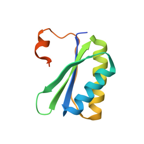
Bacterial microcompartments form a protective proteinaceous barrier around metabolic enzymes that process unstable or toxic chemical intermediates. The genome of the virulent, multidrug-resistant Clostridium difficile 630 strain contains an operon, eut, encoding a bacterial microcompartment with genes for the breakdown of ethanolamine and its utilisation as a source of reduced nitrogen and carbon. The C. difficile eut operon displays regulatory genetic elements and protein encoding regions in common with homologous loci found in the genomes of other bacteria, including the enteric pathogens Salmonella enterica and Enterococcus faecalis. The crystal structures of two microcompartment shell proteins, CD1908 and CD1918, and an uncharacterised protein with potential enzymatic activity, CD1925, were determined by X-ray crystallography. CD1908 and CD1918 display the same protein fold, though the order of secondary structure elements is permuted in CD1908 and this protein displays an N-terminal β-strand extension. These proteins form hexamers with molecules related by crystallographic and non-crystallographic symmetry. The structure of CD1925 has a cupin β-barrel fold and a putative active site that is distinct from the metal-ion dependent catalytic cupins. Thin-section transmission electron microscopy of Escherichia coli over-expressing eut proteins indicates that CD1918 is capable of self-association into arrays, suggesting an organisational role for CD1918 in the formation of this microcompartment. The work presented provides the basis for further study of the architecture and function of the C. difficile eut microcompartment, its role in metabolism and the wider consequences of intestinal colonisation and virulence in this pathogen.
- Institute for Cell and Molecular Biosciences, Newcastle University, Newcastle upon Tyne, United Kingdom.
Organizational Affiliation: