4AX9

Human thrombin complexed with Napsagatran, RO0466240


Experimental Data Snapshot

  • Method: X-RAY DIFFRACTION
  • Resolution: 1.90 Å
  • R-Value Free: 0.196 
  • R-Value Work: 0.161 
  • R-Value Observed: 0.163 

wwPDB Validation   3D Report Full Report


Ligand Structure Quality Assessment 


This is version 1.3 of the entry. See complete history


Literature

Design and Synthesis of Potent and Highly Selective Thrombin Inhibitors.

Hilpert, K.Ackermann, J.Banner, D.W.Gast, A.Gubernator, K.Hadvary, P.Labler, L.Muller, K.Schmid, G.Tschopp, T.B.

(1994) J Med Chem 37: 3889

  • DOI: https://doi.org/10.1021/jm00049a008
  • Primary Citation of Related Structures:  
    4AX9, 4AYV, 4AYY, 4AZ2

  • PubMed Abstract: 

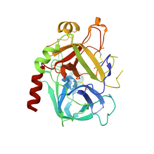
    Thrombin, a serine protease, plays a central role in the initiation and propagation of thrombotic events. An extensive search for new thrombin inhibitors was performed, using an unconventional approach. Screening of small basic molecules for binding in the recognition pocket of thrombin led to the discovery of (aminoiminomethyl)piperidine (amidinopiperidine) as a weak, but intrinsically selective, thrombin inhibitor. Elaboration of this molecule provided compounds which inhibit thrombin with Ki's in the range of 20-50 nM and with selectivities of 1000-4000 against trypsin. These inhibitor compounds show a new and unexpected binding mode to thrombin. Modification of the central building block and then of one of the hydrophobic substituents led to the discovery of a new family of thrombin inhibitors which has reverted to the former binding mode to thrombin. This last class of compounds shows inhibitory activities in the picomolar range, low toxicity, and a short plasma half life which favors its use for an intravenous application. From this series of thrombin inhibitors, 19f(Ro 46-6240) was selected for clinical development as an antithrombotic agent for intravenous administration.


  • Organizational Affiliation

    Pharma Division, F. Hoffmann-La Roche Ltd., Basel, Switzerland.


Macromolecules
Find similar proteins by:  (by identity cutoff)  |  3D Structure
Entity ID: 1
MoleculeChains Sequence LengthOrganismDetailsImage
PROTHROMBINA [auth H]257Homo sapiensMutation(s): 0 
EC: 3.4.21.5
UniProt & NIH Common Fund Data Resources
Find proteins for P00734 (Homo sapiens)
Explore P00734 
Go to UniProtKB:  P00734
PHAROS:  P00734
GTEx:  ENSG00000180210 
Entity Groups  
Sequence Clusters30% Identity50% Identity70% Identity90% Identity95% Identity100% Identity
UniProt GroupP00734
Sequence Annotations
Expand
  • Reference Sequence

Find similar proteins by:  Sequence   |   3D Structure  

Entity ID: 2
MoleculeChains Sequence LengthOrganismDetailsImage
HIRUDIN VARIANT-1B [auth I]11Hirudo medicinalisMutation(s): 0 
UniProt
Find proteins for P01050 (Hirudo medicinalis)
Explore P01050 
Go to UniProtKB:  P01050
Entity Groups  
Sequence Clusters30% Identity50% Identity70% Identity90% Identity95% Identity100% Identity
UniProt GroupP01050
Sequence Annotations
Expand
  • Reference Sequence
Find similar proteins by:  (by identity cutoff)  |  3D Structure
Entity ID: 3
MoleculeChains Sequence LengthOrganismDetailsImage
PROTHROMBINC [auth L]28Homo sapiensMutation(s): 0 
EC: 3.4.21.5
UniProt & NIH Common Fund Data Resources
Find proteins for P00734 (Homo sapiens)
Explore P00734 
Go to UniProtKB:  P00734
PHAROS:  P00734
GTEx:  ENSG00000180210 
Entity Groups  
Sequence Clusters30% Identity50% Identity70% Identity90% Identity95% Identity100% Identity
UniProt GroupP00734
Sequence Annotations
Expand
  • Reference Sequence
Small Molecules
Ligands 3 Unique
IDChains Name / Formula / InChI Key2D Diagram3D Interactions
N5N
Query on N5N

Download Ideal Coordinates CCD File 
F [auth H]2-[[(2S)-4-[[(3S)-1-carbamimidoylpiperidin-3-yl]methylamino]-2-(naphthalen-2-ylsulfonylamino)-4-oxidanylidene-butanoyl] -cyclopropyl-amino]ethanoic acid
C26 H34 N6 O6 S
BYDKEYCXCIVOOV-JTSKRJEESA-N
NAG
Query on NAG

Download Ideal Coordinates CCD File 
G [auth H]2-acetamido-2-deoxy-beta-D-glucopyranose
C8 H15 N O6
OVRNDRQMDRJTHS-FMDGEEDCSA-N
NA
Query on NA

Download Ideal Coordinates CCD File 
D [auth H],
E [auth H]
SODIUM ION
Na
FKNQFGJONOIPTF-UHFFFAOYSA-N
Binding Affinity Annotations 
IDSourceBinding Affinity
N5N Binding MOAD:  4AX9 Ki: 0.27 (nM) from 1 assay(s)
PDBBind:  4AX9 Ki: 0.27 (nM) from 1 assay(s)
BindingDB:  4AX9 Ki: min: 0.27, max: 0.3 (nM) from 2 assay(s)
Experimental Data & Validation

Experimental Data

  • Method: X-RAY DIFFRACTION
  • Resolution: 1.90 Å
  • R-Value Free: 0.196 
  • R-Value Work: 0.161 
  • R-Value Observed: 0.163 
  • Space Group: C 1 2 1
Unit Cell:
Length ( Å )Angle ( ˚ )
a = 71.4α = 90
b = 72.04β = 100.8
c = 72.7γ = 90
Software Package:
Software NamePurpose
REFMACrefinement
XDSdata reduction
XDSdata scaling
AMoREphasing

Structure Validation

View Full Validation Report


Ligand Structure Quality Assessment 


Entry History 

Deposition Data

Revision History  (Full details and data files)

  • Version 1.0: 2012-06-20
    Type: Initial release
  • Version 1.1: 2013-08-07
    Changes: Other
  • Version 1.2: 2015-09-16
    Changes: Source and taxonomy
  • Version 1.3: 2020-07-29
    Type: Remediation
    Reason: Carbohydrate remediation
    Changes: Data collection, Derived calculations, Other, Structure summary