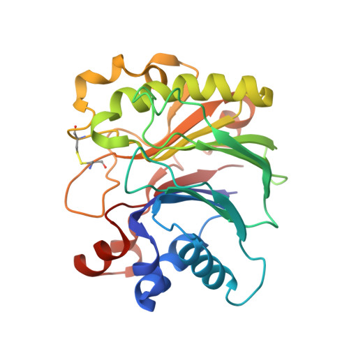
4AWN

Structure of recombinant human DNase I (rhDNaseI) in complex with Magnesium and Phosphate.


Experimental Data Snapshot

  • Method: X-RAY DIFFRACTION
  • Resolution: 1.95 Å
  • R-Value Free: 0.218 
  • R-Value Work: 0.166 
  • R-Value Observed: 0.169 

Starting Model: experimental
View more details

wwPDB Validation   3D Report Full Report


Ligand Structure Quality Assessment 


This is version 2.1 of the entry. See complete history


Literature

The Structure of Human DNase I Bound to Magnesium and Phosphate Ions Points to a Catalytic Mechanism Common to Members of the DNase I-Like Superfamily.

Parsiegla, G.Noguere, C.Santell, L.Lazarus, R.A.Bourne, Y.

(2012) Biochemistry 51: 10250

  • DOI: https://doi.org/10.1021/bi300873f
  • Primary Citation of Related Structures:  
    4AWN

  • PubMed Abstract: 

    Recombinant human DNase I (Pulmozyme, dornase alfa) is used for the treatment of cystic fibrosis where it improves lung function and reduces the number of exacerbations. The physiological mechanism of action is thought to involve the reduction of the viscoelasticity of cystic fibrosis sputum by hydrolyzing high concentrations of DNA into low-molecular mass fragments. Here we describe the 1.95 Å resolution crystal structure of recombinant human DNase I (rhDNase I) in complex with magnesium and phosphate ions, both bound in the active site. Complementary mutagenesis data of rhDNase I coupled to a comprehensive structural analysis of the DNase I-like superfamily argue for the key catalytic role of Asn7, which is invariant among mammalian DNase I enzymes and members of this superfamily, through stabilization of the magnesium ion coordination sphere. Overall, our combined structural and mutagenesis data suggest the occurrence of a magnesium-assisted pentavalent phosphate transition state in human DNase I during catalysis, where Asp168 may play a key role as a general catalytic base.


  • Organizational Affiliation

    Architecture et Fonction des Macromolécules Biologiques, Aix-Marseille Université and CNRS UMR 7257, Parc Scientifique et Technonlogique de Luminy, Case 932, 163 Avenue de Luminy, 13288 Marseille cedex 09, France. goetz.parsiegla@imm.cnrs.fr


Macromolecules
Find similar proteins by:  (by identity cutoff)  |  3D Structure
Entity ID: 1
MoleculeChains Sequence LengthOrganismDetailsImage
DEOXYRIBONUCLEASE-1260Homo sapiensMutation(s): 0 
EC: 3.1.21.1
UniProt & NIH Common Fund Data Resources
Find proteins for P24855 (Homo sapiens)
Explore P24855 
Go to UniProtKB:  P24855
PHAROS:  P24855
GTEx:  ENSG00000213918 
Entity Groups  
Sequence Clusters30% Identity50% Identity70% Identity90% Identity95% Identity100% Identity
UniProt GroupP24855
Glycosylation
Glycosylation Sites: 2Go to GlyGen: P24855-1
Sequence Annotations
Expand
  • Reference Sequence
Oligosaccharides

Help

Entity ID: 2
MoleculeChains Length2D Diagram Glycosylation3D Interactions
2-acetamido-2-deoxy-beta-D-glucopyranose-(1-4)-2-acetamido-2-deoxy-beta-D-glucopyranose
B
2N-Glycosylation
Glycosylation Resources
GlyTouCan:  G42666HT
GlyCosmos:  G42666HT
GlyGen:  G42666HT
Small Molecules
Ligands 5 Unique
IDChains Name / Formula / InChI Key2D Diagram3D Interactions
NAG
Query on NAG

Download Ideal Coordinates CCD File 
H [auth A]2-acetamido-2-deoxy-beta-D-glucopyranose
C8 H15 N O6
OVRNDRQMDRJTHS-FMDGEEDCSA-N
BTB
Query on BTB

Download Ideal Coordinates CCD File 
G [auth A]2-[BIS-(2-HYDROXY-ETHYL)-AMINO]-2-HYDROXYMETHYL-PROPANE-1,3-DIOL
C8 H19 N O5
OWMVSZAMULFTJU-UHFFFAOYSA-N
PO4
Query on PO4

Download Ideal Coordinates CCD File 
F [auth A]PHOSPHATE ION
O4 P
NBIIXXVUZAFLBC-UHFFFAOYSA-K
CA
Query on CA

Download Ideal Coordinates CCD File 
C [auth A],
D [auth A]
CALCIUM ION
Ca
BHPQYMZQTOCNFJ-UHFFFAOYSA-N
MG
Query on MG

Download Ideal Coordinates CCD File 
E [auth A]MAGNESIUM ION
Mg
JLVVSXFLKOJNIY-UHFFFAOYSA-N
Experimental Data & Validation

Experimental Data

  • Method: X-RAY DIFFRACTION
  • Resolution: 1.95 Å
  • R-Value Free: 0.218 
  • R-Value Work: 0.166 
  • R-Value Observed: 0.169 
  • Space Group: P 21 21 21
Unit Cell:
Length ( Å )Angle ( ˚ )
a = 41.96α = 90
b = 58.66β = 90
c = 96.43γ = 90
Software Package:
Software NamePurpose
REFMACrefinement
XDSdata reduction
SCALEPACKdata scaling
MOLREPphasing

Structure Validation

View Full Validation Report


Ligand Structure Quality Assessment 


Entry History 

Deposition Data

Revision History  (Full details and data files)

  • Version 1.0: 2013-01-09
    Type: Initial release
  • Version 1.1: 2013-01-23
    Changes: Database references
  • Version 1.2: 2019-05-08
    Changes: Data collection, Derived calculations, Experimental preparation, Other
  • Version 2.0: 2020-07-29
    Type: Remediation
    Reason: Carbohydrate remediation
    Changes: Atomic model, Data collection, Derived calculations, Other, Structure summary
  • Version 2.1: 2023-12-20
    Changes: Data collection, Database references, Refinement description, Structure summary