4AWJ | pdb_00004awj

pVHL:EloB:EloC complex, in complex with capped Hydroxyproline


Experimental Data Snapshot

  • Method: X-RAY DIFFRACTION
  • Resolution: 2.50 Å
  • R-Value Free: 
    0.264 (Depositor), 0.261 (DCC) 
  • R-Value Work: 
    0.220 (Depositor), 0.220 (DCC) 
  • R-Value Observed: 
    0.222 (Depositor) 

wwPDB Validation 3D Report Full Report

Validation slider image for 4AWJ

Ligand Structure Quality Assessment 


This is version 1.1 of the entry. See complete history

Literature

Dissecting Fragment-Based Lead Discovery at the Von Hippel-Lindau Protein:Hypoxia Inducible Factor 1Alpha Protein-Protein Interface.

Van Molle, I.Thomann, A.Buckley, D.L.So, E.C.Lang, S.Crews, C.M.Ciulli, A.

(2012) Chem Biol 19: 1300

  • DOI: https://doi.org/10.1016/j.chembiol.2012.08.015
  • Primary Citation Related Structures: 
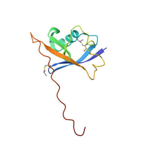
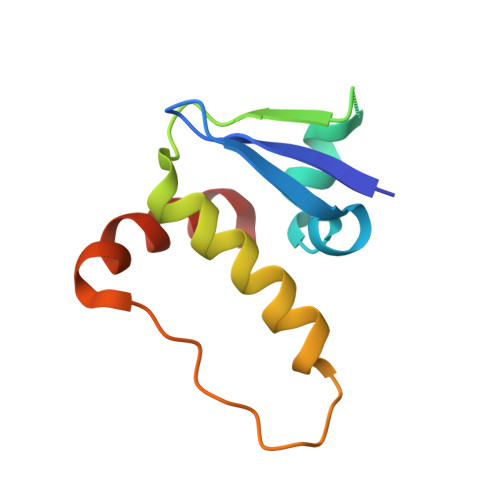
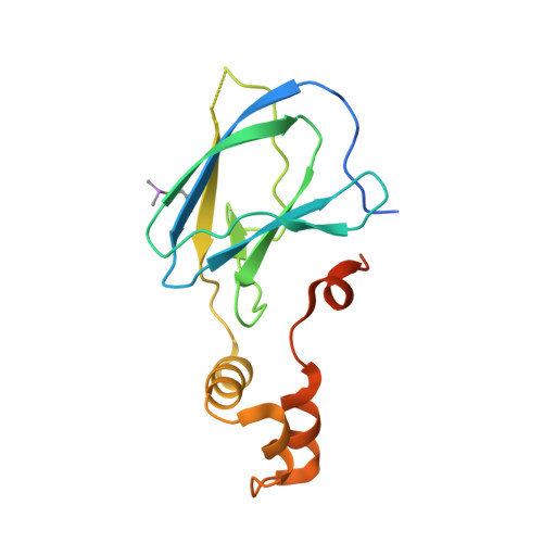
    3ZTC, 3ZTD, 4AJY, 4AWJ

  • PubMed Abstract: 

    Fragment screening is widely used to identify attractive starting points for drug design. However, its potential and limitations to assess the tractability of often challenging protein:protein interfaces have been underexplored. Here, we address this question by means of a systematic deconstruction of lead-like inhibitors of the pVHL:HIF-1α interaction into their component fragments. Using biophysical techniques commonly employed for screening, we could only detect binding of fragments that violate the Rule of Three, are more complex than those typically screened against classical druggable targets, and occupy two adjacent binding subsites at the interface rather than just one. Analyses based on ligand and group lipophilicity efficiency of anchored fragments were applied to dissect the individual subsites and probe for binding hot spots. The implications of our findings for targeting protein interfaces by fragment-based approaches are discussed.


  • Organizational Affiliation
    • Department of Chemistry, University of Cambridge, Cambridge, CB2 1EW, UK.

Macromolecule Content 

  • Total Structure Weight: 168.75 kDa 
  • Atom Count: 10,786 
  • Modeled Residue Count: 1,320 
  • Deposited Residue Count: 1,456 
  • Unique protein chains: 3

Macromolecules

Find similar proteins by:|  3D Structure
Entity ID: 1
MoleculeChains  Sequence LengthOrganismDetailsImage
TRANSCRIPTION ELONGATION FACTOR B POLYPEPTIDE 2
A, D, G, J
104Homo sapiensMutation(s): 0 
UniProt & NIH Common Fund Data Resources
Find proteins for Q15370 (Homo sapiens)
Explore Q15370 
Go to UniProtKB:  Q15370
PHAROS:  Q15370
GTEx:  ENSG00000103363 
Entity Groups
Sequence Clusters30% Identity50% Identity70% Identity90% Identity95% Identity100% Identity
UniProt GroupQ15370
Sequence Annotations
Expand
Reference Sequence
Find similar proteins by:|  3D Structure
Entity ID: 2
MoleculeChains  Sequence LengthOrganismDetailsImage
TRANSCRIPTION ELONGATION FACTOR B POLYPEPTIDE 1
B, E, H, K
97Homo sapiensMutation(s): 0 
UniProt & NIH Common Fund Data Resources
Find proteins for Q15369 (Homo sapiens)
Explore Q15369 
Go to UniProtKB:  Q15369
PHAROS:  Q15369
GTEx:  ENSG00000154582 
Entity Groups
Sequence Clusters30% Identity50% Identity70% Identity90% Identity95% Identity100% Identity
UniProt GroupQ15369
Sequence Annotations
Expand
Reference Sequence
Find similar proteins by:|  3D Structure
Entity ID: 3
MoleculeChains  Sequence LengthOrganismDetailsImage
VON HIPPEL-LINDAU DISEASE TUMOR SUPPRESSOR
C, F, I, L
163Homo sapiensMutation(s): 0 
UniProt & NIH Common Fund Data Resources
Find proteins for P40337 (Homo sapiens)
Explore P40337 
Go to UniProtKB:  P40337
PHAROS:  P40337
GTEx:  ENSG00000134086 
Entity Groups
Sequence Clusters30% Identity50% Identity70% Identity90% Identity95% Identity100% Identity
UniProt GroupP40337
Sequence Annotations
Expand
Reference Sequence

Small Molecules

Ligands 4 Unique
IDChains Name / Formula / InChI Key2D Diagram3D Interactions
V6F

Query on V6F


Download:Ideal Coordinates CCD File
O [auth C],
Q [auth F],
T [auth I],
W [auth L]
(4R)-1-acetyl-4-hydroxy-N-methyl-L-prolinamide
C8 H14 N2 O3
XAZYBLFYZNUKHD-RQJHMYQMSA-N
GOL

Query on GOL


Download:Ideal Coordinates CCD File
V [auth J]GLYCEROL
C3 H8 O3
PEDCQBHIVMGVHV-UHFFFAOYSA-N
ACY

Query on ACY


Download:Ideal Coordinates CCD File
R [auth F]ACETIC ACID
C2 H4 O2
QTBSBXVTEAMEQO-UHFFFAOYSA-N
ACT

Query on ACT


Download:Ideal Coordinates CCD File
M [auth A]
N [auth B]
P [auth C]
S [auth F]
U [auth I]
M [auth A],
N [auth B],
P [auth C],
S [auth F],
U [auth I],
X [auth L]
ACETATE ION
C2 H3 O2
QTBSBXVTEAMEQO-UHFFFAOYSA-M
Modified Residues  1 Unique
IDChains TypeFormula2D DiagramParent
CAS
Query on CAS
A, D, G, J
L-PEPTIDE LINKINGC5 H12 As N O2 SCYS
Binding Affinity Annotations 
IDSourceBinding Affinity
V6F BindingDB:  4AWJ Kd: min: 4.90e+6, max: 1.00e+7 (nM) from 2 assay(s)

Experimental Data & Validation

Experimental Data

  • Method: X-RAY DIFFRACTION
  • Resolution: 2.50 Å
  • R-Value Free:  0.264 (Depositor), 0.261 (DCC) 
  • R-Value Work:  0.220 (Depositor), 0.220 (DCC) 
  • R-Value Observed: 0.222 (Depositor) 
Space Group: P 41 2 2
Unit Cell:
Length ( Å )Angle ( ˚ )
a = 93.4α = 90
b = 93.4β = 90
c = 362.04γ = 90
Software Package:
Software NamePurpose
PHENIXrefinement
XDSdata reduction
XSCALEdata scaling
PHENIXphasing

Structure Validation

View Full Validation Report


Ligand Structure Quality Assessment 


Entry History 

Deposition Data

Revision History  (Full details and data files)

  • Version 1.0: 2012-11-14
    Type: Initial release
  • Version 1.1: 2024-11-06
    Changes: Data collection, Database references, Derived calculations, Other, Structure summary