4AUJ | pdb_00004auj

FimH lectin domain co-crystal with a alpha-D-mannoside O-linked to para hydroxypropargyl phenyl


Experimental Data Snapshot

  • Method: X-RAY DIFFRACTION
  • Resolution: 1.53 Å
  • R-Value Free: 
    0.185 (Depositor), 0.178 (DCC) 
  • R-Value Work: 
    0.160 (Depositor), 0.155 (DCC) 
  • R-Value Observed: 
    0.162 (Depositor) 

Starting Model: experimental
View more details

wwPDB Validation 3D Report Full Report

Validation slider image for 4AUJ

Ligand Structure Quality Assessment 


This is version 1.6 of the entry. See complete history

Literature

Validation of Reactivity Descriptors to Assess the Aromatic Stacking within the Tyrosine Gate of Fimh.

Roos, G.Wellens, A.Touaibia, M.Yamakawa, N.Geerlings, P.Roy, R.Wyns, L.Bouchaert, J.

(2013) ACS Med Chem Lett 4: 1085

  • DOI: https://doi.org/10.1021/ml400269v
  • Primary Citation Related Structures: 
    4ATT, 4AUJ, 4BUQ

  • PubMed Abstract: 

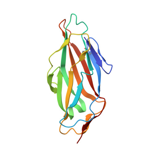
    Antagonists of the FimH adhesin, a protein almost universally present at the extremity of type-1 fimbriae expressed by Escherichia coli, have been abundantly in the spotlight as alternative treatments of urinary tract infections. The antagonists function as bacterial antiadhesives through highly specific α-d-mannose binding in a charged and polar pocket at the tip of the FimH lectin domain and by the stacking of alkyl or aromatic moieties substituted on the mannose with two tyrosine residues (Tyr48 and Tyr137) at the entrance of the mannose-binding pocket. Using high-resolution crystal data, interaction energies are calculated for the different observed aromatic stacking modes between the tyrosines and the antagonist. The dispersion component of the interaction energy correlates with the observed electron density. The quantum chemical reactivity descriptors local hardness and polarizability were successfully validated as prediction tools for ligand affinity in the tyrosine gate of FimH and therefore have potential for rapid drug screening.


  • Organizational Affiliation
    • General Chemistry, Vrije Universiteit Brussel, Structural Biology Brussels, VIB and Vrije Universiteit Brussel, and Brussels Center for Redox Biology, Pleinlaan 2, 1050 Brussels, Belgium ; General Chemistry, Vrije Universiteit Brussel, Structural Biology Brussels, VIB and Vrije Universiteit Brussel, and Brussels Center for Redox Biology, Pleinlaan 2, 1050 Brussels, Belgium ; General Chemistry, Vrije Universiteit Brussel, Structural Biology Brussels, VIB and Vrije Universiteit Brussel, and Brussels Center for Redox Biology, Pleinlaan 2, 1050 Brussels, Belgium.

Macromolecule Content 

  • Total Structure Weight: 17.23 kDa 
  • Atom Count: 1,452 
  • Modeled Residue Count: 158 
  • Deposited Residue Count: 158 
  • Unique protein chains: 1

Macromolecules

Find similar proteins by:|  3D Structure
Entity ID: 1
MoleculeChains  Sequence LengthOrganismDetailsImage
FIMH158Escherichia coli UTI89Mutation(s): 0 
UniProt
Find proteins for P08191 (Escherichia coli (strain K12))
Explore P08191 
Go to UniProtKB:  P08191
Entity Groups
Sequence Clusters30% Identity50% Identity70% Identity90% Identity95% Identity100% Identity
UniProt GroupP08191
Sequence Annotations
Expand
Reference Sequence

Small Molecules

Ligands 1 Unique
IDChains Name / Formula / InChI Key2D Diagram3D Interactions
HNW

Query on HNW


Download:Ideal Coordinates CCD File
B [auth A]4-(3-hydroxyprop-1-yn-1-yl)phenyl alpha-D-mannopyranoside
C15 H18 O7
GNTPQQUJBAFHOR-MRLBHPIUSA-N

Experimental Data & Validation

Experimental Data

  • Method: X-RAY DIFFRACTION
  • Resolution: 1.53 Å
  • R-Value Free:  0.185 (Depositor), 0.178 (DCC) 
  • R-Value Work:  0.160 (Depositor), 0.155 (DCC) 
  • R-Value Observed: 0.162 (Depositor) 
Space Group: P 21 21 21
Unit Cell:
Length ( Å )Angle ( ˚ )
a = 32.92α = 90
b = 42.29β = 90
c = 96.34γ = 90
Software Package:
Software NamePurpose
PHENIXrefinement
XDSdata reduction
XSCALEdata scaling
PHASERphasing

Structure Validation

View Full Validation Report


Ligand Structure Quality Assessment 


Entry History 

Deposition Data

Revision History  (Full details and data files)

  • Version 1.0: 2013-05-29
    Type: Initial release
  • Version 1.1: 2013-12-25
    Changes: Database references
  • Version 1.2: 2014-06-18
    Changes: Database references
  • Version 1.3: 2020-07-29
    Type: Remediation
    Reason: Carbohydrate remediation
    Changes: Derived calculations, Other, Structure summary
  • Version 1.4: 2023-12-20
    Changes: Data collection, Database references, Refinement description, Structure summary
  • Version 1.5: 2024-05-08
    Changes: Data collection
  • Version 1.6: 2024-10-23
    Changes: Structure summary