4ATP

Structure of GABA-transaminase A1R958 from Arthrobacter aurescens in complex with PLP


Experimental Data Snapshot

  • Method: X-RAY DIFFRACTION
  • Resolution: 2.80 Å
  • R-Value Free: 0.229 
  • R-Value Work: 0.193 
  • R-Value Observed: 0.195 

wwPDB Validation   3D Report Full Report


Ligand Structure Quality Assessment 


This is version 1.2 of the entry. See complete history


Literature

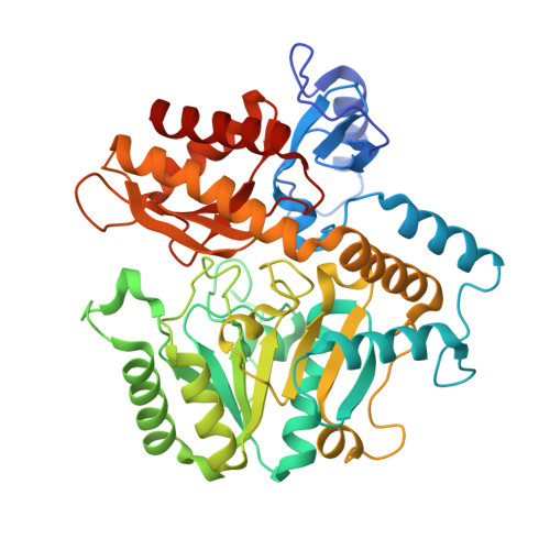
Structures of a Gamma-Aminobutyrate (Gaba) Transaminase from the S-Triazine-Degrading Organism Arthrobacter Aurescens Tc1 in Complex with Plp and with its External Aldimine Plp- Gaba Adduct.

Bruce, H.Nguyen Tuan, A.Mangas Sanchez, J.Leese, C.Hopwood, J.Hyde, R.Hart, S.Turkenburg, J.P.Grogan, G.

(2012) Acta Crystallogr Sect F Struct Biol Cryst Commun 68: 1175

  • DOI: https://doi.org/10.1107/S1744309112030023
  • Primary Citation of Related Structures:  
    4ATP, 4ATQ

  • PubMed Abstract: 

    Two complex structures of the γ-aminobutyrate (GABA) transaminase A1R958 from Arthrobacter aurescens TC1 are presented. The first, determined to a resolution of 2.80 Å, features the internal aldimine formed by reaction between the ℇ-amino group of Lys295 and the cofactor pyridoxal phosphate (PLP); the second, determined to a resolution of 2.75 Å, features the external aldimine adduct formed between PLP and GABA in the first half-reaction. This is the first structure of a microbial GABA transaminase in complex with its natural external aldimine and reveals the molecular determinants of GABA binding in this enzyme.


  • Organizational Affiliation

    York Structural Biology Laboratory, Department of Chemistry, University of York, Heslington, York YO10 5DD, England.


Macromolecules
Find similar proteins by:  (by identity cutoff)  |  3D Structure
Entity ID: 1
MoleculeChains Sequence LengthOrganismDetailsImage
4-AMINOBUTYRATE TRANSAMINASE
A, B, C, D, E
A, B, C, D, E, F, G, H, I, J, K, L
456Paenarthrobacter aurescens TC1Mutation(s): 0 
EC: 2.6.1.19
UniProt
Find proteins for A1R958 (Paenarthrobacter aurescens (strain TC1))
Explore A1R958 
Go to UniProtKB:  A1R958
Entity Groups  
Sequence Clusters30% Identity50% Identity70% Identity90% Identity95% Identity100% Identity
UniProt GroupA1R958
Sequence Annotations
Expand
  • Reference Sequence
Small Molecules
Ligands 1 Unique
IDChains Name / Formula / InChI Key2D Diagram3D Interactions
PLP
Query on PLP

Download Ideal Coordinates CCD File 
M [auth A]
N [auth B]
O [auth C]
P [auth D]
Q [auth E]
M [auth A],
N [auth B],
O [auth C],
P [auth D],
Q [auth E],
R [auth F],
S [auth G],
T [auth H],
U [auth I],
V [auth J],
W [auth K],
X [auth L]
PYRIDOXAL-5'-PHOSPHATE
C8 H10 N O6 P
NGVDGCNFYWLIFO-UHFFFAOYSA-N
Experimental Data & Validation

Experimental Data

  • Method: X-RAY DIFFRACTION
  • Resolution: 2.80 Å
  • R-Value Free: 0.229 
  • R-Value Work: 0.193 
  • R-Value Observed: 0.195 
  • Space Group: P 21 21 2
Unit Cell:
Length ( Å )Angle ( ˚ )
a = 175.7α = 90
b = 290.99β = 90
c = 104.54γ = 90
Software Package:
Software NamePurpose
REFMACrefinement
xia2data scaling
MOLREPphasing

Structure Validation

View Full Validation Report


Ligand Structure Quality Assessment 


Entry History 

Deposition Data

Revision History  (Full details and data files)

  • Version 1.0: 2012-10-10
    Type: Initial release
  • Version 1.1: 2012-10-17
    Changes: Database references
  • Version 1.2: 2023-12-20
    Changes: Data collection, Database references, Derived calculations, Other, Refinement description