4AS2

Pseudomonas Aeruginosa Phosphorylcholine Phosphatase. Monoclinic form


Experimental Data Snapshot

  • Method: X-RAY DIFFRACTION
  • Resolution: 2.12 Å
  • R-Value Free: 0.210 
  • R-Value Work: 0.154 
  • R-Value Observed: 0.157 

wwPDB Validation   3D Report Full Report


This is version 1.3 of the entry. See complete history


Literature

The Structural Domains of Pseudomonas Aeruginosa Phosphorylcholine Phosphatase Cooperate in Substrate Hydrolysis: 3D Structure and Enzymatic Mechanism.

Infantes, L.Otero, L.H.Beassoni, P.R.Boetsch, C.Lisa, A.T.Domenech, C.E.Albert, A.

(2012) J Mol Biol 423: 503

  • DOI: https://doi.org/10.1016/j.jmb.2012.07.024
  • Primary Citation of Related Structures:  
    4AS2, 4AS3

  • PubMed Abstract: 

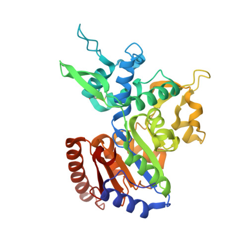
    Pseudomonas aeruginosa is an opportunistic Gram-negative pathogen. It colonizes different tissues by the utilization of diverse mechanisms. One of these may involve the breakdown of the host cell membrane through the sequential action of hemolytic phospholipase C and phosphorylcholine phosphatase (PchP). The action of hemolytic phospholipase C on phosphatidylcholine produces phosphorylcholine, which is hydrolyzed to choline (Cho) and inorganic phosphate by PchP. The available biochemical data on this enzyme demonstrate the involvement of two Cho-binding sites in the catalytic cycle and in enzyme regulation. The crystal structure of P. aeruginosa PchP has been determined. It folds into three structural domains. The first domain harbors all the residues involved in catalysis and is well conserved among the haloacid dehalogenase superfamily of proteins. The second domain is characteristic of PchP and is involved in the recognition of the Cho moiety of the substrate. The third domain stabilizes the relative position of the other two. Fortuitously, the crystal structure of PchP captures molecules of Bistris (2-[bis(2-hydroxyethyl)amino]-2-(hydroxymethyl)propane-1,3-diol) at the active site and at an additional site. This represents two catalytically relevant complexes with just one or two inhibitory Bistris molecules and provides the basis of the PchP function and regulation. Site-directed mutagenesis along with biochemical experiments corroborates the structural observations and demonstrates the interplay between different sites for Cho recognition and inhibition. The structural comparison of PchP with other phosphatases of the haloacid dehalogenase family provides a three-dimensional picture of the conserved catalytic cycle and the structural basis for the recognition of the diverse substrate molecules.


  • Organizational Affiliation

    Departamento de Cristalografía y Biología Estructural, Instituto de Química Física Rocasolano, CSIC, Madrid, Spain.


Macromolecules
Find similar proteins by:  (by identity cutoff)  |  3D Structure
Entity ID: 1
MoleculeChains Sequence LengthOrganismDetailsImage
PHOSPHORYLCHOLINE PHOSPHATASE
A, B, C, D
327Pseudomonas aeruginosaMutation(s): 0 
EC: 3.1.3.75
UniProt
Find proteins for Q9HTR2 (Pseudomonas aeruginosa (strain ATCC 15692 / DSM 22644 / CIP 104116 / JCM 14847 / LMG 12228 / 1C / PRS 101 / PAO1))
Explore Q9HTR2 
Go to UniProtKB:  Q9HTR2
Entity Groups  
Sequence Clusters30% Identity50% Identity70% Identity90% Identity95% Identity100% Identity
UniProt GroupQ9HTR2
Sequence Annotations
Expand
  • Reference Sequence
Small Molecules
Ligands 4 Unique
IDChains Name / Formula / InChI Key2D Diagram3D Interactions
BTB
Query on BTB

Download Ideal Coordinates CCD File 
AA [auth C]
GA [auth D]
J [auth A]
K [auth A]
L [auth A]
AA [auth C],
GA [auth D],
J [auth A],
K [auth A],
L [auth A],
M [auth A],
R [auth B],
Z [auth C]
2-[BIS-(2-HYDROXY-ETHYL)-AMINO]-2-HYDROXYMETHYL-PROPANE-1,3-DIOL
C8 H19 N O5
OWMVSZAMULFTJU-UHFFFAOYSA-N
IOD
Query on IOD

Download Ideal Coordinates CCD File 
DA [auth D]
EA [auth D]
FA [auth D]
G [auth A]
H [auth A]
DA [auth D],
EA [auth D],
FA [auth D],
G [auth A],
H [auth A],
I [auth A],
N [auth A],
Q [auth B],
S [auth B],
V [auth C],
W [auth C],
X [auth C],
Y [auth C]
IODIDE ION
I
XMBWDFGMSWQBCA-UHFFFAOYSA-M
CL
Query on CL

Download Ideal Coordinates CCD File 
CA [auth D],
F [auth A],
P [auth B],
U [auth C]
CHLORIDE ION
Cl
VEXZGXHMUGYJMC-UHFFFAOYSA-M
MG
Query on MG

Download Ideal Coordinates CCD File 
BA [auth D],
E [auth A],
O [auth B],
T [auth C]
MAGNESIUM ION
Mg
JLVVSXFLKOJNIY-UHFFFAOYSA-N
Experimental Data & Validation

Experimental Data

  • Method: X-RAY DIFFRACTION
  • Resolution: 2.12 Å
  • R-Value Free: 0.210 
  • R-Value Work: 0.154 
  • R-Value Observed: 0.157 
  • Space Group: C 1 2 1
Unit Cell:
Length ( Å )Angle ( ˚ )
a = 136.006α = 90
b = 156.633β = 118.03
c = 71.816γ = 90
Software Package:
Software NamePurpose
REFMACrefinement
XDSdata reduction
SCALEdata scaling
SHELXphasing

Structure Validation

View Full Validation Report


Entry History 

Deposition Data

Revision History  (Full details and data files)

  • Version 1.0: 2012-08-22
    Type: Initial release
  • Version 1.1: 2012-09-26
    Changes: Database references
  • Version 1.2: 2012-10-31
    Changes: Database references
  • Version 1.3: 2019-05-08
    Changes: Data collection, Experimental preparation, Other