Calcium-Dependent Complex Formation between Pbp2 and Lytic Transglycosylase Sltb1 of Pseudomonas Aeruginosa.
Nikolaidis, I., Izore, T., Job, V., Thielens, N., Breukink, E., Dessen, A.(2012) Microb Drug Resist 18: 298
- PubMed: 22432706 Search on PubMed
- DOI: https://doi.org/10.1089/mdr.2012.0006
- Primary Citation Related Structures:
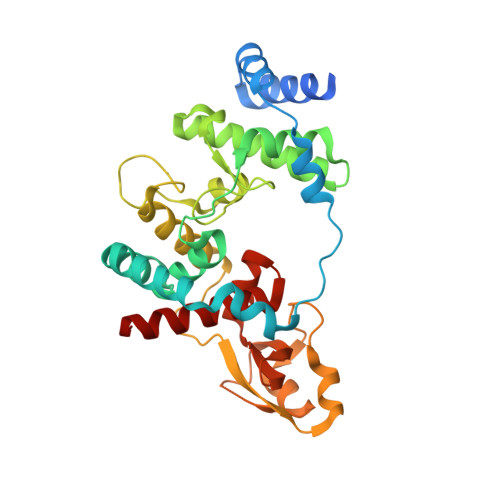
4ANR - PubMed Abstract:
In Gram-negative bacteria, the bacterial cell wall biosynthetic mechanism requires the coordinated action of enzymes and structural proteins located in the cytoplasm, within the membrane, and in the periplasm of the cell. Its main component, peptidoglycan (PG), is essential for cell division and wall elongation. Penicillin-binding proteins (PBPs) catalyze the last steps of PG biosynthesis, namely the polymerization of glycan chains and the cross-linking of stem peptides, and can be either monofunctional or bifunctional. Their action is coordinated with that of other enzymes essential for cell-wall biosynthesis, such as lytic transglycosylases (LT). Here, we have studied SltB1, an LT from Pseudomonas aeruginosa, and identified that it forms a complex with PBP2, a monofunctional enzyme, which requires the presence of Ca(2+). In addition, we have solved the structure of SltB1 to a high resolution, and identified that it harbors an EF-hand like motif containing a Ca(2+) ion displaying bipyramidal coordination. These studies provide initial structural details that shed light on the interactions between the PG biosynthesis enzymes in P. aeruginosa.
- Bacterial Pathogenesis Group, Institut de Biologie Structurale (IBS), Université Grenoble I, Grenoble, France.
Organizational Affiliation: