4AIN | pdb_00004ain

Crystal structure of BetP with asymmetric protomers.


Experimental Data Snapshot

  • Method: X-RAY DIFFRACTION
  • Resolution: 3.10 Å
  • R-Value Free: 
    0.285 (Depositor), 0.286 (DCC) 
  • R-Value Work: 
    0.225 (Depositor), 0.226 (DCC) 
  • R-Value Observed: 
    0.229 (Depositor) 

wwPDB Validation 3D Report Full Report

Validation slider image for 4AIN

Ligand Structure Quality Assessment 


This is version 2.0 of the entry. See complete history

Literature

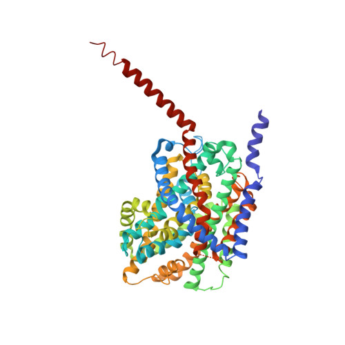
Alternating-Access Mechanism in Conformationally Asymmetric Trimers of the Betaine Transporter Betp.

Perez, C.Koshy, C.Yildiz, O.Ziegler, C.

(2012) Nature 490: 126

  • DOI: https://doi.org/10.1038/nature11403
  • Primary Citation Related Structures: 
    4AIN, 4DOJ

  • PubMed Abstract: 

    Betaine and Na(+) symport has been extensively studied in the osmotically regulated transporter BetP from Corynebacterium glutamicum, a member of the betaine/choline/carnitine transporter family, which shares the conserved LeuT-like fold of two inverted structural repeats. BetP adjusts its transport activity by sensing the cytoplasmic K(+) concentration as a measure for hyperosmotic stress via the osmosensing carboxy-terminal domain. BetP needs to be in a trimeric state for communication between individual protomers through several intratrimeric interaction sites. Recently, crystal structures of inward-facing BetP trimers have contributed to our understanding of activity regulation on a molecular level. Here we report new crystal structures, which reveal two conformationally asymmetric BetP trimers, capturing among them three distinct transport states. We observe a total of four new conformations at once: an outward-open apo and an outward-occluded apo state, and two closed transition states--one in complex with betaine and one substrate-free. On the basis of these new structures, we identified local and global conformational changes in BetP that underlie the molecular transport mechanism, which partially resemble structural changes observed in other sodium-coupled LeuT-like fold transporters, but show differences we attribute to the osmolytic nature of betaine, the exclusive substrate specificity and the regulatory properties of BetP.


  • Organizational Affiliation
    • Max-Planck Institute of Biophysics, Department of Structural Biology, D-60438 Frankfurt am Main, Germany.

Macromolecule Content 

  • Total Structure Weight: 175.47 kDa 
  • Atom Count: 11,639 
  • Modeled Residue Count: 1,525 
  • Deposited Residue Count: 1,617 
  • Unique protein chains: 1

Macromolecules

Find similar proteins by:|  3D Structure
Entity ID: 1
MoleculeChains  Sequence LengthOrganismDetailsImage
GLYCINE BETAINE TRANSPORTER BETP
A, B, C
539Corynebacterium glutamicumMutation(s): 0 
Membrane Entity: Yes 
UniProt
Find proteins for P54582 (Corynebacterium glutamicum (strain ATCC 13032 / DSM 20300 / JCM 1318 / BCRC 11384 / CCUG 27702 / LMG 3730 / NBRC 12168 / NCIMB 10025 / NRRL B-2784 / 534))
Explore P54582 
Go to UniProtKB:  P54582
Entity Groups
Sequence Clusters30% Identity50% Identity70% Identity90% Identity95% Identity100% Identity
UniProt GroupP54582
Sequence Annotations
Expand
Reference Sequence

Small Molecules

Ligands 6 Unique
IDChains Name / Formula / InChI Key2D Diagram3D Interactions
CM5

Query on CM5


Download:Ideal Coordinates CCD File
M [auth C]5-CYCLOHEXYL-1-PENTYL-BETA-D-MALTOSIDE
C23 H42 O11
RVTGFZGNOSKUDA-ZNGNCRBCSA-N
FLC

Query on FLC


Download:Ideal Coordinates CCD File
L [auth C]CITRATE ANION
C6 H5 O7
KRKNYBCHXYNGOX-UHFFFAOYSA-K
BET

Query on BET


Download:Ideal Coordinates CCD File
E [auth B],
J [auth C]
TRIMETHYL GLYCINE
C5 H12 N O2
KWIUHFFTVRNATP-UHFFFAOYSA-O
PEG

Query on PEG


Download:Ideal Coordinates CCD File
F [auth B]DI(HYDROXYETHYL)ETHER
C4 H10 O3
MTHSVFCYNBDYFN-UHFFFAOYSA-N
CL

Query on CL


Download:Ideal Coordinates CCD File
G [auth B],
H [auth C],
I [auth C],
K [auth C]
CHLORIDE ION
Cl
VEXZGXHMUGYJMC-UHFFFAOYSA-M
NA

Query on NA


Download:Ideal Coordinates CCD File
D [auth B]SODIUM ION
Na
FKNQFGJONOIPTF-UHFFFAOYSA-N

Experimental Data & Validation

Experimental Data

  • Method: X-RAY DIFFRACTION
  • Resolution: 3.10 Å
  • R-Value Free:  0.285 (Depositor), 0.286 (DCC) 
  • R-Value Work:  0.225 (Depositor), 0.226 (DCC) 
  • R-Value Observed: 0.229 (Depositor) 
Space Group: P 21 21 21
Unit Cell:
Length ( Å )Angle ( ˚ )
a = 112.81α = 90
b = 128.15β = 90
c = 159.69γ = 90
Software Package:
Software NamePurpose
PHENIXrefinement
XDSdata reduction
XSCALEdata scaling

Structure Validation

View Full Validation Report


Ligand Structure Quality Assessment 


Entry History 

Deposition Data

Revision History  (Full details and data files)

  • Version 1.0: 2012-06-06
    Type: Initial release
  • Version 1.1: 2012-08-29
    Changes: Database references
  • Version 1.2: 2012-09-12
    Changes: Database references
  • Version 1.3: 2012-10-10
    Changes: Database references
  • Version 1.4: 2013-03-27
    Changes: Other
  • Version 1.5: 2015-03-25
    Changes: Database references
  • Version 2.0: 2023-11-15
    Changes: Atomic model, Data collection, Database references, Derived calculations, Other