4ACJ

Crystal structure of the TLDC domain of Oxidation resistance protein 2 from zebrafish


Experimental Data Snapshot

  • Method: X-RAY DIFFRACTION
  • Resolution: 0.97 Å
  • R-Value Free: 0.138 
  • R-Value Work: 0.119 
  • R-Value Observed: 0.119 

wwPDB Validation   3D Report Full Report


This is version 1.2 of the entry. See complete history


Literature

Crystal Structure of the Tldc Domain of Oxidation Resistance Protein 2 from Zebrafish.

Blaise, M.Alsarraf, H.M.Wong, J.E.Midtgaard, S.R.Laroche, F.Schack, L.Spaink, H.Stougaard, J.Thirup, S.

(2012) Proteins 80: 1694

  • DOI: https://doi.org/10.1002/prot.24050
  • Primary Citation of Related Structures:  
    4ACJ

  • PubMed Abstract: 

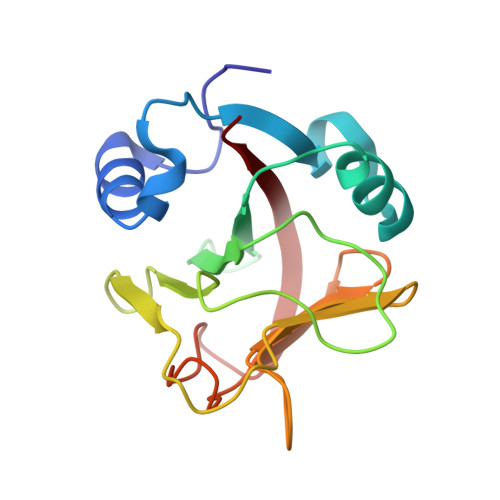
    The oxidation resistance proteins (OXR) help to protect eukaryotes from reactive oxygen species. The sole C-terminal domain of the OXR, named TLDc is sufficient to perform this function. However, the mechanism by which oxidation resistance occurs is poorly understood. We present here the crystal structure of the TLDc domain of the oxidation resistance protein 2 from zebrafish. The structure was determined by X-ray crystallography to atomic resolution (0.97Å) and adopts an overall globular shape. Two antiparallel β-sheets form a central β-sandwich, surrounded by two helices and two one-turn helices. The fold shares low structural similarity to known structures.


  • Organizational Affiliation

    Department of Molecular Biology and Genetics, Centre for Carbohydrate Recognition and Signalling, Aarhus University, Aarhus, Denmark. mick@mb.au.dk


Macromolecules
Find similar proteins by:  (by identity cutoff)  |  3D Structure
Entity ID: 1
MoleculeChains Sequence LengthOrganismDetailsImage
WU\:FB25H12 PROTEIN,167Danio rerioMutation(s): 0 
UniProt
Find proteins for A9JTH8 (Danio rerio)
Explore A9JTH8 
Go to UniProtKB:  A9JTH8
Entity Groups  
Sequence Clusters30% Identity50% Identity70% Identity90% Identity95% Identity100% Identity
UniProt GroupA9JTH8
Sequence Annotations
Expand
  • Reference Sequence
Experimental Data & Validation

Experimental Data

  • Method: X-RAY DIFFRACTION
  • Resolution: 0.97 Å
  • R-Value Free: 0.138 
  • R-Value Work: 0.119 
  • R-Value Observed: 0.119 
  • Space Group: P 21 21 2
Unit Cell:
Length ( Å )Angle ( ˚ )
a = 65.62α = 90
b = 69.11β = 90
c = 36.27γ = 90
Software Package:
Software NamePurpose
PHENIXrefinement
PHENIXphasing

Structure Validation

View Full Validation Report


Entry History 

Deposition Data

Revision History  (Full details and data files)

  • Version 1.0: 2012-02-08
    Type: Initial release
  • Version 1.1: 2012-05-23
    Changes: Other
  • Version 1.2: 2018-01-17
    Changes: Advisory, Data collection