Crystallographic Study of Novel Transthyretin Ligands Exhibiting Negative-Cooperativity between Two Thyroxine Binding Sites.
Tomar, D., Khan, T., Singh, R.R., Mishra, S., Gupta, S., Surolia, A., Salunke, D.M.(2012) PLoS One 7: 43522
- PubMed: 22973437 Search on PubMedSearch on PubMed Central
- DOI: https://doi.org/10.1371/journal.pone.0043522
- Primary Citation Related Structures:
4ABQ, 4ABU, 4ABV, 4ABW, 4AC2, 4AC4, 4ANK - PubMed Abstract:
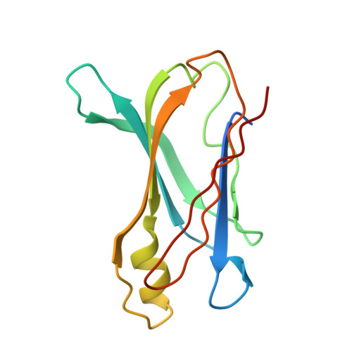
Transthyretin (TTR) is a homotetrameric serum and cerebrospinal fluid protein that transports thyroxine (T4) and retinol by binding to retinol binding protein. Rate-limiting tetramer dissociation and rapid monomer misfolding and disassembly of TTR lead to amyloid fibril formation in different tissues causing various amyloid diseases. Based on the current understanding of the pathogenesis of TTR amyloidosis, it is considered that the inhibition of amyloid fibril formation by stabilization of TTR in native tetrameric form is a viable approach for the treatment of TTR amyloidosis. We have examined interactions of the wtTTR with a series of compounds containing various substitutions at biphenyl ether skeleton and a novel compound, previously evaluated for binding and inhibiting tetramer dissociation, by x-ray crystallographic approach. High resolution crystal structures of five ligands in complex with wtTTR provided snapshots of negatively cooperative binding of ligands in two T4 binding sites besides characterizing their binding orientations, conformations, and interactions with binding site residues. In all complexes, the ligand has better fit and more potent interactions in first T4 site i.e. (AC site) than the second T4 site (BD site). Together, these results suggest that AC site is a preferred ligand binding site and retention of ordered water molecules between the dimer interfaces further stabilizes the tetramer by bridging a hydrogen bond interaction between Ser117 and its symmetric copy. Novel biphenyl ether based compounds exhibit negative-cooperativity while binding to two T4 sites which suggests that binding of only single ligand molecule is sufficient to inhibit the TTR tetramer dissociation.
- Structural Biology Unit, National Institute of Immunology, New Delhi, India.
Organizational Affiliation: