4AAW | pdb_00004aaw

S.pneumoniae GlmU in complex with an antibacterial inhibitor


Experimental Data Snapshot

  • Method: X-RAY DIFFRACTION
  • Resolution: 2.20 Å
  • R-Value Free: 
    0.237 (Depositor), 0.260 (DCC) 
  • R-Value Work: 
    0.200 (Depositor) 
  • R-Value Observed: 
    0.202 (Depositor) 

Starting Model: experimental
View more details

wwPDB Validation 3D Report Full Report

Validation slider image for 4AAW

Ligand Structure Quality Assessment 


This is version 1.3 of the entry. See complete history

Literature

Inhibitors of Acetyltransferase Domain of N-Acetylglucosamine-1-Phosphate-Uridyltransferase/ Glucosamine-1-Phosphate-Acetyltransferase (Glmu). Part 1: Hit to Lead Evaluation of a Novel Arylsulfonamide Series.

Green, O.M.McKenzie, A.R.Shapiro, A.B.Otterbein, L.Ni, H.Patten, A.Stokes, S.Albert, R.Kawatkar, S.Breed, J.

(2012) Bioorg Med Chem Lett 22: 1510

  • DOI: https://doi.org/10.1016/j.bmcl.2012.01.016
  • Primary Citation Related Structures: 
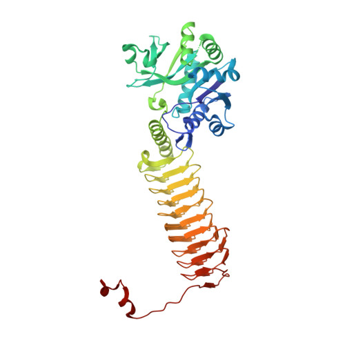
    4AA7, 4AAW, 4AC3

  • PubMed Abstract: 

    A novel arylsulfonamide-containing series of compounds represented by 1, discovered by highthroughput screening, inhibit the acetyltransferase domain of N-acetylglucosamine-1-phosphate-uridyltransferase/glucosamine-1-phosphate-acetyltransferase (GlmU). X-ray structure determination confirmed that inhibitor binds at the site occupied by acetyl-CoA, indicating that series is competitive with this substrate. This letter documents our early hit-to-lead evaluation of the chemical series and some of the findings that led to improvement in in-vitro potency against Gram-negative and Gram-positive bacterial isozymes, exemplified by compound 40.


  • Organizational Affiliation
    • Infection Innovative Medicines Unit, AstraZeneca R&D Boston, Waltham, MA 02451, USA. oluyinka.green@astrazeneca.com

Macromolecule Content 

  • Total Structure Weight: 50.21 kDa 
  • Atom Count: 3,790 
  • Modeled Residue Count: 455 
  • Deposited Residue Count: 459 
  • Unique protein chains: 1

Macromolecules

Find similar proteins by:|  3D Structure
Entity ID: 1
MoleculeChains  Sequence LengthOrganismDetailsImage
BIFUNCTIONAL PROTEIN GLMU459Streptococcus pneumoniaeMutation(s): 0 
EC: 2.3.1.157 (PDB Primary Data), 2.7.7.23 (PDB Primary Data)
UniProt
Find proteins for Q8DQ18 (Streptococcus pneumoniae (strain ATCC BAA-255 / R6))
Explore Q8DQ18 
Go to UniProtKB:  Q8DQ18
Entity Groups
Sequence Clusters30% Identity50% Identity70% Identity90% Identity95% Identity100% Identity
UniProt GroupQ8DQ18
Sequence Annotations
Expand
Reference Sequence

Small Molecules

Ligands 2 Unique
IDChains Name / Formula / InChI Key2D Diagram3D Interactions
R84

Query on R84


Download:Ideal Coordinates CCD File
B [auth A]4-{[1-(2-{[({5-[(3-carboxypropanoyl)amino]-2,4-dimethoxyphenyl}sulfonyl)amino]methyl}phenyl)piperidin-4-yl]methoxy}-4-oxobutanoic acid
C29 H37 N3 O11 S
PRDHHOYRFMTLDT-UHFFFAOYSA-N
SO4

Query on SO4


Download:Ideal Coordinates CCD File
C [auth A]SULFATE ION
O4 S
QAOWNCQODCNURD-UHFFFAOYSA-L
Binding Affinity Annotations 
IDSourceBinding Affinity
R84 BindingDB:  4AAW IC50: 500 (nM) from 1 assay(s)

Experimental Data & Validation

Experimental Data

  • Method: X-RAY DIFFRACTION
  • Resolution: 2.20 Å
  • R-Value Free:  0.237 (Depositor), 0.260 (DCC) 
  • R-Value Work:  0.200 (Depositor) 
  • R-Value Observed: 0.202 (Depositor) 
Space Group: P 3 2 1
Unit Cell:
Length ( Å )Angle ( ˚ )
a = 116.779α = 90
b = 116.779β = 90
c = 116.683γ = 120
Software Package:
Software NamePurpose
REFMACrefinement
d*TREKdata reduction
d*TREKdata scaling
AMoREphasing

Structure Validation

View Full Validation Report


Ligand Structure Quality Assessment 


Entry History 

Deposition Data

Revision History  (Full details and data files)

  • Version 1.0: 2012-02-15
    Type: Initial release
  • Version 1.1: 2012-08-15
    Changes: Database references
  • Version 1.2: 2017-12-27
    Changes: Database references
  • Version 1.3: 2023-12-20
    Changes: Data collection, Database references, Derived calculations, Other, Refinement description