4A9A

Structure of Rbg1 in complex with Tma46 dfrp domain


Experimental Data Snapshot

  • Method: X-RAY DIFFRACTION
  • Resolution: 2.67 Å
  • R-Value Free: 0.222 
  • R-Value Work: 0.196 
  • R-Value Observed: 0.197 

wwPDB Validation   3D Report Full Report


This is version 1.2 of the entry. See complete history


Literature

Rbg1-Tma46 Dimer Structure Reveals New Functional Domains and Their Role in Polysome Recruitment.

Francis, S.M.Gas, M.Daugeron, M.Bravo, J.Seraphin, B.

(2012) Nucleic Acids Res 40: 11100

  • DOI: https://doi.org/10.1093/nar/gks867
  • Primary Citation of Related Structures:  
    4A9A

  • PubMed Abstract: 

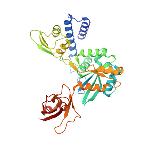
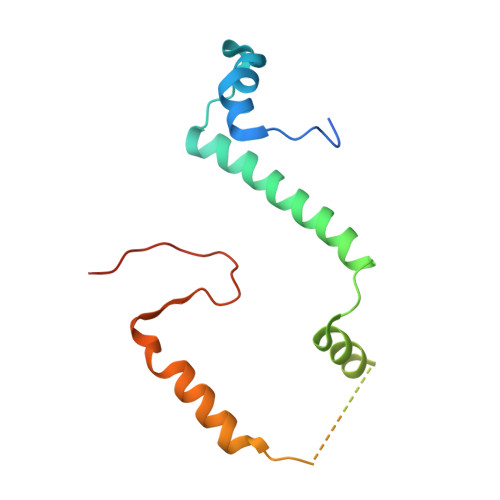
    Developmentally Regulated GTP-binding (DRG) proteins are highly conserved GTPases that associate with DRG Family Regulatory Proteins (DFRP). The resulting complexes have recently been shown to participate in eukaryotic translation. The structure of the Rbg1 GTPase, a yeast DRG protein, in complex with the C-terminal region of its DFRP partner, Tma46, was solved by X-ray diffraction. These data reveal that DRG proteins are multimodular factors with three additional domains, helix-turn-helix (HTH), S5D2L and TGS, packing against the GTPase platform. Surprisingly, the S5D2L domain is inserted in the middle of the GTPase sequence. In contrast, the region of Tma46 interacting with Rbg1 adopts an extended conformation typical of intrinsically unstructured proteins and contacts the GTPase and TGS domains. Functional analyses demonstrate that the various domains of Rbg1, as well as Tma46, modulate the GTPase activity of Rbg1 and contribute to the function of these proteins in vivo. Dissecting the role of the different domains revealed that the Rbg1 TGS domain is essential for the recruitment of this factor in polysomes, supporting further the implication of these conserved factors in translation.


  • Organizational Affiliation

    Instituto de Biomedicina de Valencia (IBV-CSIC), Calle Jaime Roig, 11, Valencia E-46010, Spain.


Macromolecules
Find similar proteins by:  (by identity cutoff)  |  3D Structure
Entity ID: 1
MoleculeChains Sequence LengthOrganismDetailsImage
RIBOSOME-INTERACTING GTPASE 1
A, B
376Saccharomyces cerevisiaeMutation(s): 0 
UniProt
Find proteins for P39729 (Saccharomyces cerevisiae (strain ATCC 204508 / S288c))
Explore P39729 
Go to UniProtKB:  P39729
Entity Groups  
Sequence Clusters30% Identity50% Identity70% Identity90% Identity95% Identity100% Identity
UniProt GroupP39729
Sequence Annotations
Expand
  • Reference Sequence
Find similar proteins by:  (by identity cutoff)  |  3D Structure
Entity ID: 2
MoleculeChains Sequence LengthOrganismDetailsImage
TRANSLATION MACHINERY-ASSOCIATED PROTEIN 46
C, D
142Saccharomyces cerevisiaeMutation(s): 0 
UniProt
Find proteins for Q12000 (Saccharomyces cerevisiae (strain ATCC 204508 / S288c))
Explore Q12000 
Go to UniProtKB:  Q12000
Entity Groups  
Sequence Clusters30% Identity50% Identity70% Identity90% Identity95% Identity100% Identity
UniProt GroupQ12000
Sequence Annotations
Expand
  • Reference Sequence
Experimental Data & Validation

Experimental Data

  • Method: X-RAY DIFFRACTION
  • Resolution: 2.67 Å
  • R-Value Free: 0.222 
  • R-Value Work: 0.196 
  • R-Value Observed: 0.197 
  • Space Group: P 21 21 2
Unit Cell:
Length ( Å )Angle ( ˚ )
a = 86.2α = 90
b = 224.89β = 90
c = 84.89γ = 90
Software Package:
Software NamePurpose
REFMACrefinement
MOSFLMdata reduction
SCALAdata scaling
SHARPphasing

Structure Validation

View Full Validation Report


Entry History 

Deposition Data

Revision History  (Full details and data files)

  • Version 1.0: 2012-10-03
    Type: Initial release
  • Version 1.1: 2012-12-19
    Changes: Database references
  • Version 1.2: 2017-01-25
    Changes: Data collection