4A92 | pdb_00004a92

Full-length HCV NS3-4A protease-helicase in complex with a macrocyclic protease inhibitor.


Experimental Data Snapshot

  • Method: X-RAY DIFFRACTION
  • Resolution: 2.73 Å
  • R-Value Free: 
    0.231 (Depositor), 0.229 (DCC) 
  • R-Value Work: 
    0.178 (Depositor), 0.178 (DCC) 
  • R-Value Observed: 
    0.181 (Depositor) 

wwPDB Validation 3D Report Full Report

Validation slider image for 4A92

Ligand Structure Quality Assessment 


This is version 1.3 of the entry. See complete history

Literature

A Macrocyclic Hcv Ns3/4A Protease Inhibitor Interacts with Protease and Helicase Residues in the Complex with its Full- Length Target.

Schiering, N.D'Arcy, A.Villard, F.Simic, O.Kamke, M.Monnet, G.Hassiepen, U.Svergun, D.I.Pulfer, R.Eder, J.Raman, P.Bodendorf, U.

(2011) Proc Natl Acad Sci U S A 108: 21052

  • DOI: https://doi.org/10.1073/pnas.1110534108
  • Primary Citation Related Structures: 
    4A92

  • PubMed Abstract: 

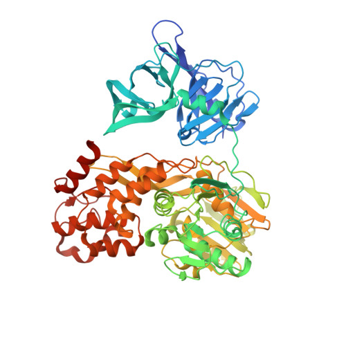
    Hepatitis C virus (HCV) infection is a global health burden with over 170 million people infected worldwide. In a significant portion of patients chronic hepatitis C infection leads to serious liver diseases, including fibrosis, cirrhosis, and hepatocellular carcinoma. The HCV NS3 protein is essential for viral polyprotein processing and RNA replication and hence viral replication. It is composed of an N-terminal serine protease domain and a C-terminal helicase/NTPase domain. For full activity, the protease requires the NS4A protein as a cofactor. HCV NS3/4A protease is a prime target for developing direct-acting antiviral agents. First-generation NS3/4A protease inhibitors have recently been introduced into clinical practice, markedly changing HCV treatment options. To date, crystal structures of HCV NS3/4A protease inhibitors have only been reported in complex with the protease domain alone. Here, we present a unique structure of an inhibitor bound to the full-length, bifunctional protease-helicase NS3/4A and show that parts of the P4 capping and P2 moieties of the inhibitor interact with both protease and helicase residues. The structure sheds light on inhibitor binding to the more physiologically relevant form of the enzyme and supports exploring inhibitor-helicase interactions in the design of the next generation of HCV NS3/4A protease inhibitors. In addition, small angle X-ray scattering confirmed the observed protease-helicase domain assembly in solution.


  • Organizational Affiliation
    • Expertise Platform Proteases, Novartis Institutes for BioMedical Research, Fabrikstrasse 16, CH-4002 Basel, Switzerland. nikolaus.schiering@novartis.com

Macromolecule Content 

  • Total Structure Weight: 143.51 kDa 
  • Atom Count: 9,848 
  • Modeled Residue Count: 1,278 
  • Deposited Residue Count: 1,332 
  • Unique protein chains: 1

Macromolecules

Find similar proteins by:|  3D Structure
Entity ID: 1
MoleculeChains  Sequence LengthOrganismDetailsImage
SERINE PROTEASE NS3
A, B
666Orthohepacivirus hominisMutation(s): 2 
EC: 3.4.21.98 (PDB Primary Data), 3.6.1.15 (PDB Primary Data), 3.6.4.13 (PDB Primary Data)
UniProt
Find proteins for P26663 (Hepatitis C virus genotype 1b (isolate BK))
Explore P26663 
Go to UniProtKB:  P26663
Entity Groups
Sequence Clusters30% Identity50% Identity70% Identity90% Identity95% Identity100% Identity
UniProt GroupP26663
Sequence Annotations
Expand
Reference Sequence

Small Molecules

Ligands 2 Unique
IDChains Name / Formula / InChI Key2D Diagram3D Interactions
F9K

Query on F9K


Download:Ideal Coordinates CCD File
D [auth A],
F [auth B]
(1'R,2R,2'S,6S,24AS)-17-FLUORO-6-(1-METHYL-2-OXOPIPERIDINE-3-CARBOXAMIDO)-19,19-DIOXIDO-5,21,24-TRIOXO-2'-VINYL-1,2,3,5,6,7,8,9,10,11,12,13,14,20,21,23,24,24A-OCTADECAHYDROSPIRO[BENZO[S]PYRROLO[2,1-G][1,2,5,8,18]THIATETRAAZACYCLOICOSINE-22,1'-CYCLOPRO-2-CARBOXYLATEPAN]-2-YL 4-FLUOROISOINDOLINE
C42 H51 F2 N7 O9 S
JSGPAIBYTVNGSI-ZBBLQTOUSA-N
ZN

Query on ZN


Download:Ideal Coordinates CCD File
C [auth A],
E [auth B]
ZINC ION
Zn
PTFCDOFLOPIGGS-UHFFFAOYSA-N

Experimental Data & Validation

Experimental Data

  • Method: X-RAY DIFFRACTION
  • Resolution: 2.73 Å
  • R-Value Free:  0.231 (Depositor), 0.229 (DCC) 
  • R-Value Work:  0.178 (Depositor), 0.178 (DCC) 
  • R-Value Observed: 0.181 (Depositor) 
Space Group: P 21 21 21
Unit Cell:
Length ( Å )Angle ( ˚ )
a = 91.972α = 90
b = 110.47β = 90
c = 137.227γ = 90
Software Package:
Software NamePurpose
BUSTERrefinement
XDSdata reduction
XSCALEdata scaling

Structure Validation

View Full Validation Report


Ligand Structure Quality Assessment 


Entry History 

Deposition Data

Revision History  (Full details and data files)

  • Version 1.0: 2011-12-28
    Type: Initial release
  • Version 1.1: 2012-01-11
    Changes: Other
  • Version 1.2: 2019-05-08
    Changes: Data collection, Experimental preparation, Other
  • Version 1.3: 2024-05-08
    Changes: Data collection, Database references, Derived calculations, Other