4A7G | pdb_00004a7g

Structure of human I113T SOD1 mutant complexed with 4-methylpiperazin- 1-yl)quinazoline in the p21 space group.


Experimental Data Snapshot

  • Method: X-RAY DIFFRACTION
  • Resolution: 1.24 Å
  • R-Value Free: 
    0.175 (Depositor), 0.184 (DCC) 
  • R-Value Work: 
    0.139 (Depositor), 0.148 (DCC) 
  • R-Value Observed: 
    0.141 (Depositor) 

wwPDB Validation 3D Report Full Report

Validation slider image for 4A7G

Ligand Structure Quality Assessment 


This is version 1.6 of the entry. See complete history

Literature

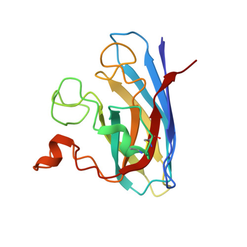
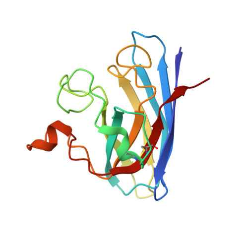
X-ray crystallography and computational docking for the detection and development of protein-ligand interactions.

Kershaw, N.M.Wright, G.S.Sharma, R.Antonyuk, S.V.Strange, R.W.Berry, N.G.O'Neill, P.M.Hasnain, S.S.

(2013) Curr Med Chem 20: 569-575

  • DOI: https://doi.org/10.2174/0929867311320040008
  • Primary Citation Related Structures: 
    4A7G, 4A7Q

  • PubMed Abstract: 

    Amyotrophic lateral sclerosis (ALS) is a progressive neurodegenerative disorder characterised by the selective dysfunction and death of the upper and lower motor neurons. Median survival rates are between 3 and 5 years after diagnosis. Mutations in the gene encoding Cu/Zn superoxide dismutase (SOD1) have been linked to a subset of familial forms of ALS (fALS). Herein, we describe a fragment- based drug discovery (FBDD) approach for the investigation of small molecule binding sites in SOD1. X-ray crystallography has been used as the primary screening method and has been shown to directly detect protein-ligand interactions which cannot be unambiguously identified using other biophysical methods. The structural requirements for effective binding at Trp32 are detailed for a series of quinazoline-containing compounds. The investigation of an additional site that binds a range of catecholamines and the use of computational modelling to assist fragment evolution is discussed. This study also highlights the importance of ligand solubility for successful Xray crystallographic campaigns in lead compound design.


  • Organizational Affiliation
    • Molecular Biophysics Group, Institute of Integrative Biology, Faculty of Health and Life Sciences, University of Liverpool, Liverpool, UK. nkershaw@liverpool.ac.uk

Macromolecule Content 

  • Total Structure Weight: 33.1 kDa 
  • Atom Count: 2,871 
  • Modeled Residue Count: 306 
  • Deposited Residue Count: 306 
  • Unique protein chains: 2

Macromolecules

Find similar proteins by:|  3D Structure
Entity ID: 1
MoleculeChains  Sequence LengthOrganismDetailsImage
SUPEROXIDE DISMUTASE [CU-ZN]153Homo sapiensMutation(s): 1 
EC: 1.15.1.1 (PDB Primary Data), 1.8 (UniProt)
UniProt & NIH Common Fund Data Resources
Find proteins for P00441 (Homo sapiens)
Explore P00441 
Go to UniProtKB:  P00441
PHAROS:  P00441
GTEx:  ENSG00000142168 
Entity Groups
Sequence Clusters30% Identity50% Identity70% Identity90% Identity95% Identity100% Identity
UniProt GroupP00441
Sequence Annotations
Expand
Reference Sequence
Find similar proteins by:|  3D Structure
Entity ID: 2
MoleculeChains  Sequence LengthOrganismDetailsImage
SUPEROXIDE DISMUTASE [CU-ZN]B [auth F]153Homo sapiensMutation(s): 1 
EC: 1.15.1.1 (PDB Primary Data), 1.8 (UniProt)
UniProt & NIH Common Fund Data Resources
Find proteins for P00441 (Homo sapiens)
Explore P00441 
Go to UniProtKB:  P00441
PHAROS:  P00441
GTEx:  ENSG00000142168 
Entity Groups
Sequence Clusters30% Identity50% Identity70% Identity90% Identity95% Identity100% Identity
UniProt GroupP00441
Sequence Annotations
Expand
Reference Sequence

Small Molecules

Ligands 5 Unique
IDChains Name / Formula / InChI Key2D Diagram3D Interactions
12I

Query on 12I


Download:Ideal Coordinates CCD File
D [auth A],
F [auth A],
O [auth F]
4-(4-METHYLPIPERAZIN-1-YL)QUINAZOLINE
C13 H16 N4
RDYFSMILBPHDCN-UHFFFAOYSA-N
SO4

Query on SO4


Download:Ideal Coordinates CCD File
C [auth A],
E [auth A],
J [auth F],
K [auth F],
M [auth F]
SULFATE ION
O4 S
QAOWNCQODCNURD-UHFFFAOYSA-L
ZN

Query on ZN


Download:Ideal Coordinates CCD File
H [auth A],
N [auth F]
ZINC ION
Zn
PTFCDOFLOPIGGS-UHFFFAOYSA-N
CU

Query on CU


Download:Ideal Coordinates CCD File
G [auth A],
L [auth F]
COPPER (II) ION
Cu
JPVYNHNXODAKFH-UHFFFAOYSA-N
ACT

Query on ACT


Download:Ideal Coordinates CCD File
I [auth A]ACETATE ION
C2 H3 O2
QTBSBXVTEAMEQO-UHFFFAOYSA-M

Experimental Data & Validation

Experimental Data

  • Method: X-RAY DIFFRACTION
  • Resolution: 1.24 Å
  • R-Value Free:  0.175 (Depositor), 0.184 (DCC) 
  • R-Value Work:  0.139 (Depositor), 0.148 (DCC) 
  • R-Value Observed: 0.141 (Depositor) 
Space Group: P 1 21 1
Unit Cell:
Length ( Å )Angle ( ˚ )
a = 38.793α = 90
b = 68.352β = 104.49
c = 49.92γ = 90
Software Package:
Software NamePurpose
REFMACrefinement
HKL-2000data reduction
HKL-2000data scaling
REFMACphasing

Structure Validation

View Full Validation Report


Ligand Structure Quality Assessment 


Entry History 

Deposition Data

Revision History  (Full details and data files)

  • Version 1.0: 2012-10-24
    Type: Initial release
  • Version 1.1: 2013-03-13
    Changes: Database references
  • Version 1.2: 2013-08-28
    Changes: Database references
  • Version 1.3: 2018-11-14
    Changes: Data collection, Database references
  • Version 1.4: 2018-11-21
    Changes: Data collection, Source and taxonomy
  • Version 1.5: 2019-10-09
    Changes: Data collection, Database references, Other
  • Version 1.6: 2024-10-16
    Changes: Data collection, Database references, Derived calculations, Structure summary