4A6A | pdb_00004a6a

A115V variant of dCTP deaminase-dUTPase from Mycobacterium tuberculosis in complex with dTTP


Experimental Data Snapshot

  • Method: X-RAY DIFFRACTION
  • Resolution: 2.90 Å
  • R-Value Free: 
    0.290 (Depositor), 0.281 (DCC) 
  • R-Value Work: 
    0.253 (Depositor), 0.246 (DCC) 
  • R-Value Observed: 
    0.255 (Depositor) 

wwPDB Validation 3D Report Full Report

Validation slider image for 4A6A

Ligand Structure Quality Assessment 


This is version 1.3 of the entry. See complete history

Literature

Dttp Inhibition of the Bifunctional Dctp Deaminase- Dutpase from Mycobacterium Tuberculosis is Ph Dependent: Kinetic Analyses and Crystal Structure of A115V Variant

Lovgreen, M.N.Harris, P.Ucar, E.Willemoes, M.

To be published.

Macromolecule Content 

  • Total Structure Weight: 257.16 kDa 
  • Atom Count: 17,912 
  • Modeled Residue Count: 2,263 
  • Deposited Residue Count: 2,280 
  • Unique protein chains: 1

Macromolecules

Find similar proteins by:|  3D Structure
Entity ID: 1
MoleculeChains  Sequence LengthOrganismDetailsImage
DEOXYCYTIDINE TRIPHOSPHATE DEAMINASE
A, B, C, D, E
A, B, C, D, E, F, G, H, I, J, K, L
190Mycobacterium tuberculosis H37RvMutation(s): 1 
EC: 3.5.4.13 (PDB Primary Data), 3.5.4.30 (UniProt)
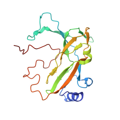
UniProt
Find proteins for P9WP17 (Mycobacterium tuberculosis (strain ATCC 25618 / H37Rv))
Explore P9WP17 
Go to UniProtKB:  P9WP17
Entity Groups
Sequence Clusters30% Identity50% Identity70% Identity90% Identity95% Identity100% Identity
UniProt GroupP9WP17
Sequence Annotations
Expand
Reference Sequence

Small Molecules

Ligands 2 Unique
IDChains Name / Formula / InChI Key2D Diagram3D Interactions
TTP

Query on TTP


Download:Ideal Coordinates CCD File
BA [auth H]
EA [auth I]
GA [auth J]
IA [auth K]
KA [auth L]
BA [auth H],
EA [auth I],
GA [auth J],
IA [auth K],
KA [auth L],
M [auth A],
O [auth B],
R [auth C],
T [auth D],
V [auth E],
X [auth F],
Z [auth G]
THYMIDINE-5'-TRIPHOSPHATE
C10 H17 N2 O14 P3
NHVNXKFIZYSCEB-XLPZGREQSA-N
MG

Query on MG


Download:Ideal Coordinates CCD File
AA [auth G]
CA [auth H]
DA [auth H]
FA [auth I]
HA [auth J]
AA [auth G],
CA [auth H],
DA [auth H],
FA [auth I],
HA [auth J],
JA [auth K],
LA [auth L],
N [auth A],
P [auth B],
Q [auth B],
S [auth C],
U [auth D],
W [auth E],
Y [auth F]
MAGNESIUM ION
Mg
JLVVSXFLKOJNIY-UHFFFAOYSA-N

Experimental Data & Validation

Experimental Data

  • Method: X-RAY DIFFRACTION
  • Resolution: 2.90 Å
  • R-Value Free:  0.290 (Depositor), 0.281 (DCC) 
  • R-Value Work:  0.253 (Depositor), 0.246 (DCC) 
  • R-Value Observed: 0.255 (Depositor) 
Space Group: P 1 21 1
Unit Cell:
Length ( Å )Angle ( ˚ )
a = 56.59α = 90
b = 179.51β = 96.28
c = 99.81γ = 90
Software Package:
Software NamePurpose
REFMACrefinement
XDSdata reduction
XSCALEdata scaling

Structure Validation

View Full Validation Report


Ligand Structure Quality Assessment 


Entry History 

Deposition Data

Revision History  (Full details and data files)

  • Version 1.0: 2012-10-10
    Type: Initial release
  • Version 1.1: 2012-12-26
    Changes: Structure summary
  • Version 1.2: 2017-11-08
    Changes: Source and taxonomy
  • Version 1.3: 2024-05-08
    Changes: Data collection, Database references, Derived calculations, Other, Refinement description