4A69 | pdb_00004a69

Structure of HDAC3 bound to corepressor and inositol tetraphosphate


Experimental Data Snapshot

  • Method: X-RAY DIFFRACTION
  • Resolution: 2.06 Å
  • R-Value Free: 
    0.236 (Depositor), 0.235 (DCC) 
  • R-Value Work: 
    0.187 (Depositor), 0.185 (DCC) 
  • R-Value Observed: 
    0.190 (Depositor) 

Starting Model: experimental
View more details

wwPDB Validation 3D Report Full Report

Validation slider image for 4A69

Ligand Structure Quality Assessment 


This is version 1.2 of the entry. See complete history

Literature

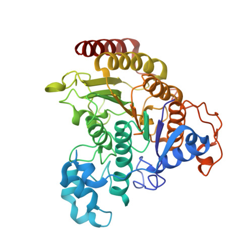
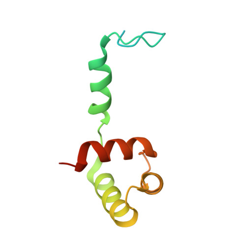
Structure of Hdac3 Bound to Co-Repressor and Inositol Tetraphosphate.

Watson, P.J.Fairall, L.Santos, G.M.Schwabe, J.W.R.

(2012) Nature 481: 335

  • DOI: https://doi.org/10.1038/nature10728
  • Primary Citation Related Structures: 
    4A69

  • PubMed Abstract: 

    Histone deacetylase enzymes (HDACs) are emerging cancer drug targets. They regulate gene expression by removing acetyl groups from lysine residues in histone tails, resulting in chromatin condensation. The enzymatic activity of most class I HDACs requires recruitment into multi-subunit co-repressor complexes, which are in turn recruited to chromatin by repressive transcription factors. Here we report the structure of a complex between an HDAC and a co-repressor, namely, human HDAC3 with the deacetylase activation domain (DAD) from the human SMRT co-repressor (also known as NCOR2). The structure reveals two remarkable features. First, the SMRT-DAD undergoes a large structural rearrangement on forming the complex. Second, there is an essential inositol tetraphosphate molecule--D-myo-inositol-(1,4,5,6)-tetrakisphosphate (Ins(1,4,5,6)P(4))--acting as an 'intermolecular glue' between the two proteins. Assembly of the complex is clearly dependent on the Ins(1,4,5,6)P(4), which may act as a regulator--potentially explaining why inositol phosphates and their kinases have been found to act as transcriptional regulators. This mechanism for the activation of HDAC3 appears to be conserved in class I HDACs from yeast to humans, and opens the way to novel therapeutic opportunities.


  • Organizational Affiliation
    • Henry Wellcome Laboratories of Structural Biology, Department of Biochemistry, University of Leicester, Leicester LE1 9HN, UK.

Macromolecule Content 

  • Total Structure Weight: 110.23 kDa 
  • Atom Count: 7,536 
  • Modeled Residue Count: 875 
  • Deposited Residue Count: 940 
  • Unique protein chains: 2

Macromolecules

Find similar proteins by:|  3D Structure
Entity ID: 1
MoleculeChains  Sequence LengthOrganismDetailsImage
HISTONE DEACETYLASE 3,
A, B
376Homo sapiensMutation(s): 0 
EC: 3.5.1.98 (PDB Primary Data), 3.5.1 (UniProt)
UniProt & NIH Common Fund Data Resources
Find proteins for O15379 (Homo sapiens)
Explore O15379 
Go to UniProtKB:  O15379
PHAROS:  O15379
GTEx:  ENSG00000171720 
Entity Groups
Sequence Clusters30% Identity50% Identity70% Identity90% Identity95% Identity100% Identity
UniProt GroupO15379
Sequence Annotations
Expand
Reference Sequence
Find similar proteins by:|  3D Structure
Entity ID: 2
MoleculeChains  Sequence LengthOrganismDetailsImage
NUCLEAR RECEPTOR COREPRESSOR 2
C, D
94Homo sapiensMutation(s): 0 
UniProt & NIH Common Fund Data Resources
Find proteins for Q9Y618 (Homo sapiens)
Explore Q9Y618 
Go to UniProtKB:  Q9Y618
PHAROS:  Q9Y618
GTEx:  ENSG00000196498 
Entity Groups
Sequence Clusters30% Identity50% Identity70% Identity90% Identity95% Identity100% Identity
UniProt GroupQ9Y618
Sequence Annotations
Expand
Reference Sequence

Small Molecules

Ligands 5 Unique
IDChains Name / Formula / InChI Key2D Diagram3D Interactions
I0P

Query on I0P


Download:Ideal Coordinates CCD File
Q [auth C],
R [auth D]
D-MYO INOSITOL 1,4,5,6 TETRAKISPHOSPHATE
C6 H16 O18 P4
MRVYFOANPDTYBY-YORTWTKJSA-N
GOL

Query on GOL


Download:Ideal Coordinates CCD File
I [auth A],
J [auth A],
O [auth B],
P [auth B]
GLYCEROL
C3 H8 O3
PEDCQBHIVMGVHV-UHFFFAOYSA-N
ZN

Query on ZN


Download:Ideal Coordinates CCD File
E [auth A],
K [auth B]
ZINC ION
Zn
PTFCDOFLOPIGGS-UHFFFAOYSA-N
ACT

Query on ACT


Download:Ideal Coordinates CCD File
F [auth A],
L [auth B]
ACETATE ION
C2 H3 O2
QTBSBXVTEAMEQO-UHFFFAOYSA-M
K

Query on K


Download:Ideal Coordinates CCD File
G [auth A],
H [auth A],
M [auth B],
N [auth B]
POTASSIUM ION
K
NPYPAHLBTDXSSS-UHFFFAOYSA-N

Experimental Data & Validation

Experimental Data

  • Method: X-RAY DIFFRACTION
  • Resolution: 2.06 Å
  • R-Value Free:  0.236 (Depositor), 0.235 (DCC) 
  • R-Value Work:  0.187 (Depositor), 0.185 (DCC) 
  • R-Value Observed: 0.190 (Depositor) 
Space Group: C 2 2 21
Unit Cell:
Length ( Å )Angle ( ˚ )
a = 86.447α = 90
b = 118.634β = 90
c = 190.707γ = 90
Software Package:
Software NamePurpose
REFMACrefinement
XDSdata reduction
SCALAdata scaling
PHASERphasing

Structure Validation

View Full Validation Report


Ligand Structure Quality Assessment 


Entry History 

Deposition Data

Revision History  (Full details and data files)

  • Version 1.0: 2012-01-11
    Type: Initial release
  • Version 1.1: 2012-01-25
    Changes: Other
  • Version 1.2: 2023-12-20
    Changes: Data collection, Database references, Derived calculations, Other, Refinement description