4A28 | pdb_00004a28

Eg5-2


Experimental Data Snapshot

  • Method: X-RAY DIFFRACTION
  • Resolution: 2.55 Å
  • R-Value Free: 
    0.264 (Depositor), 0.262 (DCC) 
  • R-Value Work: 
    0.180 (Depositor), 0.178 (DCC) 
  • R-Value Observed: 
    0.184 (Depositor) 

Starting Model: experimental
View more details

wwPDB Validation 3D Report Full Report

Validation slider image for 4A28

Ligand Structure Quality Assessment 


This is version 1.3 of the entry. See complete history

Literature

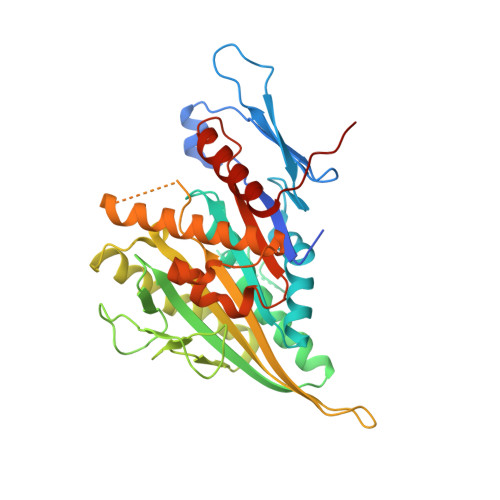
The Mitotic Kinesin Eg5 Overcomes Inhibition to the Phase I/II Clinical Candidate Sb743921 by an Allosteric Resistance Mechanism.

Talapatra, S.K.Anthony, N.G.Mackay, S.P.Kozielski, F.

(2013) J Med Chem 56: 6317

  • DOI: https://doi.org/10.1021/jm4006274
  • Primary Citation Related Structures: 
    4A1Z, 4A28, 4AS7, 4B7B, 4BXN

  • PubMed Abstract: 

    Development of drug resistance during cancer chemotherapy is one of the major causes of chemotherapeutic failure for the majority of clinical agents. The aim of this study was to investigate the underlying molecular mechanism of resistance developed by the mitotic kinesin Eg5 against the potent second-generation ispinesib analogue SB743921 (1), a phase I/II clinical candidate. Biochemical and biophysical data demonstrate that point mutations in the inhibitor-binding pocket decrease the efficacy of 1 by several 1000-fold. Surprisingly, the structures of wild-type and mutant Eg5 in complex with 1 display no apparent structural changes in the binding configuration of the drug candidate. Furthermore, ITC and modeling approaches reveal that resistance to 1 is not through conventional steric effects at the binding site but through reduced flexibility and changes in energy fluctuation pathways through the protein that influence its function. This is a phenomenon we have called "resistance by allostery".


  • Organizational Affiliation
    • The Beatson Institute for Cancer Research, Garscube Estate, Switchback Road, Bearsden, Glasgow G61 1BS, Scotland, U.K. s.talapatra@ed.ac.uk

Macromolecule Content 

  • Total Structure Weight: 83.1 kDa 
  • Atom Count: 5,306 
  • Modeled Residue Count: 654 
  • Deposited Residue Count: 736 
  • Unique protein chains: 1

Macromolecules

Find similar proteins by:|  3D Structure
Entity ID: 1
MoleculeChains  Sequence LengthOrganismDetailsImage
KINESIN-LIKE PROTEIN KIF11
A, B
368Homo sapiensMutation(s): 1 
UniProt & NIH Common Fund Data Resources
Find proteins for P52732 (Homo sapiens)
Explore P52732 
Go to UniProtKB:  P52732
PHAROS:  P52732
GTEx:  ENSG00000138160 
Entity Groups
Sequence Clusters30% Identity50% Identity70% Identity90% Identity95% Identity100% Identity
UniProt GroupP52732
Sequence Annotations
Expand
Reference Sequence

Experimental Data & Validation

Experimental Data

  • Method: X-RAY DIFFRACTION
  • Resolution: 2.55 Å
  • R-Value Free:  0.264 (Depositor), 0.262 (DCC) 
  • R-Value Work:  0.180 (Depositor), 0.178 (DCC) 
  • R-Value Observed: 0.184 (Depositor) 
Space Group: P 1 21 1
Unit Cell:
Length ( Å )Angle ( ˚ )
a = 52.9α = 90
b = 78.39β = 93.16
c = 92.69γ = 90
Software Package:
Software NamePurpose
PHENIXrefinement
MOSFLMdata reduction
SCALAdata scaling
MOLREPphasing

Structure Validation

View Full Validation Report


Ligand Structure Quality Assessment 


Entry History 

Deposition Data

Revision History  (Full details and data files)

  • Version 1.0: 2012-10-31
    Type: Initial release
  • Version 1.1: 2013-07-31
    Changes: Database references
  • Version 1.2: 2013-09-04
    Changes: Database references
  • Version 1.3: 2023-12-20
    Changes: Data collection, Database references, Derived calculations, Other, Refinement description