4A0E

Crystal structure of the cytoplasmic N-terminal domain of Yersinia pestis YscD


Experimental Data Snapshot

  • Method: X-RAY DIFFRACTION
  • Resolution: 2.04 Å
  • R-Value Free: 0.256 
  • R-Value Work: 0.211 
  • R-Value Observed: 0.213 

wwPDB Validation   3D Report Full Report


This is version 1.1 of the entry. See complete history


Literature

Structure of the Cytoplasmic Domain of Yersinia Pestis Yscd, an Essential Component of the Type III Secretion System

Lountos, G.T.Tropea, J.E.Waugh, D.S.

(2012) Acta Crystallogr D Biol Crystallogr 68: 201

  • DOI: https://doi.org/10.1107/S0907444911054308
  • Primary Citation of Related Structures:  
    4A0E

  • PubMed Abstract: 

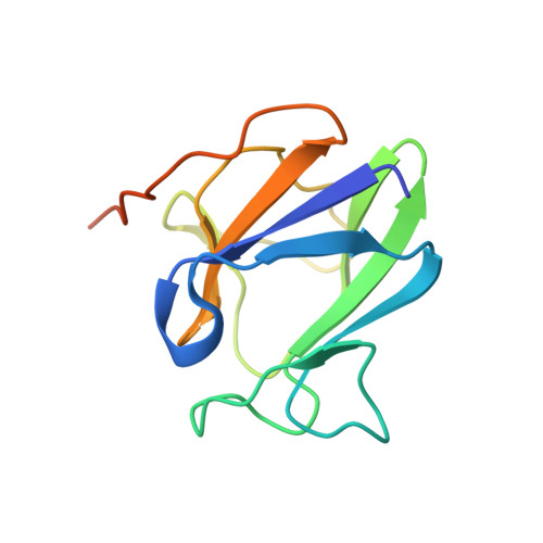
    The Yersinia pestis YscD protein is an essential component of the type III secretion system. YscD consists of an N-terminal cytoplasmic domain (residues 1-121), a transmembrane linker (122-142) and a large periplasmic domain (143-419). Both the cytoplasmic and the periplasmic domains are required for the assembly of the type III secretion system. Here, the structure of the YscD cytoplasmic domain solved by SAD phasing is presented. Although the three-dimensional structure is similar to those of forkhead-associated (FHA) domains, comparison with the structures of canonical FHA domains revealed that the cytoplasmic domain of YscD lacks the conserved residues that are required for binding phosphothreonine and is therefore unlikely to function as a true FHA domain.


  • Organizational Affiliation

    Basic Science Program, SAIC-Frederick Inc., National Cancer Institute at Frederick, Frederick, MD 21702-1201, USA.


Macromolecules
Find similar proteins by:  (by identity cutoff)  |  3D Structure
Entity ID: 1
MoleculeChains Sequence LengthOrganismDetailsImage
TYPE III SECRETION PROTEIN
A, B
123Yersinia pestisMutation(s): 0 
UniProt
Find proteins for Q56975 (Yersinia pestis)
Explore Q56975 
Go to UniProtKB:  Q56975
Entity Groups  
Sequence Clusters30% Identity50% Identity70% Identity90% Identity95% Identity100% Identity
UniProt GroupQ56975
Sequence Annotations
Expand
  • Reference Sequence
Experimental Data & Validation

Experimental Data

  • Method: X-RAY DIFFRACTION
  • Resolution: 2.04 Å
  • R-Value Free: 0.256 
  • R-Value Work: 0.211 
  • R-Value Observed: 0.213 
  • Space Group: P 4 3 2
Unit Cell:
Length ( Å )Angle ( ˚ )
a = 118.227α = 90
b = 118.227β = 90
c = 118.227γ = 90
Software Package:
Software NamePurpose
PHENIXrefinement
HKL-3000data reduction
SCALEPACKdata scaling
PHENIXphasing

Structure Validation

View Full Validation Report


Entry History 

Deposition Data

Revision History  (Full details and data files)

  • Version 1.0: 2012-02-29
    Type: Initial release
  • Version 1.1: 2012-03-28
    Changes: Other