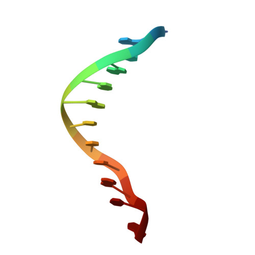
Atomic-resolution crystal structures of B-DNA reveal specific influences of divalent metal ions on conformation and packing.
Minasov, G., Tereshko, V., Egli, M.(1999) J Mol Biology 291: 83-99
- PubMed: 10438608
- DOI: https://doi.org/10.1006/jmbi.1999.2934
- Primary Citation of Related Structures:
476D, 477D, 478D - PubMed Abstract:
Crystal structures of B-form DNA have provided insights into the global and local conformational properties of the double helix, the solvent environment, drug binding and DNA packing. For example, structures of the duplex with sequence CGCGAATTCGCG, the Dickerson-Drew dodecamer (DDD), established a unique geometry of the central A-tract and a hydration spine in the minor groove. However, our knowledge of the various interaction modes between metal ions and DNA is very limited and almost no information exists concerning the origins of the different effects on DNA conformation and packing exerted by individual metal ions. Crystallization of the DDD duplex in the presence of Mg(2+)and Ca(2+)yields different crystal forms. The structures of the new Ca(2+)-form and isomorphous structures of oligonucleotides with sequences GGCGAATTCGCG and GCGAATTCGCG were determined at a maximum resolution of 1.3 A. These and the 1.1 A structure of the DDD Mg(2+)-form have revealed the most detailed picture yet of the ionic environment of B-DNA. In the Mg(2+)and Ca(2+)-forms, duplexes in the crystal lattice are surrounded by 13 magnesium and 11 calcium ions, respectively.Mg(2+)and Ca(2+)generate different DNA crystal lattices and stabilize different end-to-end overlaps and lateral contacts between duplexes, thus using different strategies for reducing the effective repeat length of the helix to ten base-pairs. Mg(2+)crystals allow the two outermost base-pairs at either end to interact laterally via minor groove H-bonds, turning the 12-mer into an effective 10-mer. Ca(2+)crystals, in contrast, unpair the outermost base-pair at each end, converting the helix into a 10-mer that can stack along its axis. This reduction of a 12-mer into a functional 10-mer is followed no matter what the detailed nature of the 5'-end of the chain: C-G-C-G-A-ellipsis, G-G-C-G-A-ellipsis, or a truncated G-C-G-A-ellipsis Rather than merely mediating close contacts between phosphate groups, ions are at the origin of many well-known features of the DDD duplex structure. A Mg(2+)coordinates in the major groove, contributing to kinking of the duplex at one end. While Ca(2+)resides in the minor groove, coordinating to bases via its hydration shell, two magnesium ions are located at the periphery of the minor groove, bridging phosphate groups from opposite strands and contracting the groove at one border of the A-tract.
- Department of Molecular Pharmacology and Biological Chemistry and The Drug Discovery Program, Northwestern University Medical School, Chicago, IL, 60611-3008, USA.
Organizational Affiliation:


