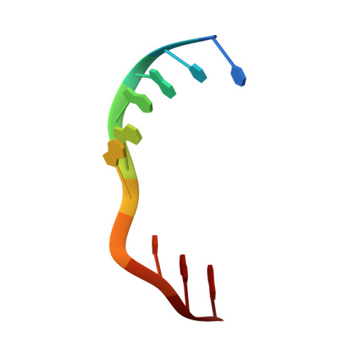
Crystal structure of a DNA Holliday junction
Ortiz-Lombardia, M., Gonzalez, A., Eritja, R., Aymami, J., Azorin, F., Coll, M.(1999) Nat Struct Biol 6: 913-917
- PubMed: 10504723 Search on PubMed
- DOI: https://doi.org/10.1038/13277
- Primary Citation Related Structures:
467D - PubMed Abstract:
DNA recombination is a universal biological event responsible both for the generation of genetic diversity and for the maintenance of genome integrity. A four-way DNA junction, also termed Holliday junction, is the key intermediate in nearly all recombination processes. This junction is the substrate of recombination enzymes that promote branch migration or catalyze its resolution. We have determined the crystal structure of a four-way DNA junction by multiwavelength anomalous diffraction, and refined it to 2.16 A resolution. The structure has two-fold symmetry, with pairwise stacking of the double-helical arms, which form two continuous B-DNA helices that run antiparallel, cross in a right-handed way, and contain two G-A mismatches. The exchanging backbones form a compact structure with strong van der Waals contacts and hydrogen bonds, implying that a conformational change must occur for the junction to branch-migrate or isomerize. At the branch point, two phosphate groups from one helix occupy the major groove of the other one, establishing sequence-specific hydrogen bonds. These interactions, together with different stacking energies and steric hindrances, explain the preference for a particular junction stacked conformer.
- Institut de Biologia Molecular de Barcelona, C.S.I.C., Jordi Girona 18, E-08034 Barcelona, Spain.
Organizational Affiliation: