407D | pdb_0000407d

STRUCTURAL BASIS FOR RECOGNITION OF A-T AND T-A BASE PAIRS IN THE MINOR GROOVE OF B-DNA


Experimental Data Snapshot

  • Method: X-RAY DIFFRACTION
  • Resolution: 2.20 Å
  • R-Value Free: 
    0.236 (Depositor) 
  • R-Value Work: 
    0.210 (Depositor) 
  • R-Value Observed: 
    0.210 (Depositor) 

wwPDB Validation 3D Report Full Report

Validation slider image for 407D

This is version 2.1 of the entry. See complete history

Literature

A structural basis for recognition of A.T and T.A base pairs in the minor groove of B-DNA.

Kielkopf, C.L.White, S.Szewczyk, J.W.Turner, J.M.Baird, E.E.Dervan, P.B.Rees, D.C.

(1998) Science 282: 111-115

  • DOI: https://doi.org/10.1126/science.282.5386.111
  • Primary Citation Related Structures: 
    407D, 408D

  • PubMed Abstract: 

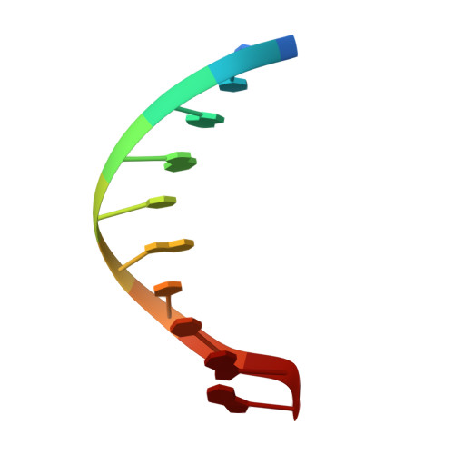
    Polyamide dimers containing three types of aromatic rings-pyrrole, imidazole, and hydroxypyrrole-afford a small-molecule recognition code that discriminates among all four Watson-Crick base pairs in the minor groove. The crystal structure of a specific polyamide dimer-DNA complex establishes the structural basis for distinguishing T.A from A.T base pairs. Specificity for the T.A base pair is achieved by means of distinct hydrogen bonds between pairs of substituted pyrroles on the ligand and the O2 of thymine and N3 of adenine. In addition, shape-selective recognition of an asymmetric cleft between the thymine-O2 and the adenine-C2 was observed. Although hitherto similarities among the base pairs in the minor groove have been emphasized, the structure illustrates differences that allow specific minor groove recognition.


  • Organizational Affiliation
    • Division of Biology, California Institute of Technology, Pasadena, CA 91125, USA.

Macromolecule Content 

  • Total Structure Weight: 7.54 kDa 
  • Atom Count: 593 
  • Modeled Residue Count: 20 
  • Deposited Residue Count: 20 
  • Unique nucleic acid chains: 1

Macromolecules

Find similar nucleic acids by:  Sequence
Entity ID: 1
MoleculeChains LengthOrganismImage
DNA (5'-D(*CP*CP*AP*GP*TP*AP*CP*TP*GP*G)-3')
A, B
10synthetic construct
Sequence Annotations
Expand
Reference Sequence

Small Molecules

Ligands 2 Unique
IDChains Name / Formula / InChI Key2D Diagram3D Interactions
HP1

Query on HP1


Download:Ideal Coordinates CCD File
C [auth A],
E [auth B]
~{N}-[5-[[5-[[5-[[3-[3-(dimethylamino)propylamino]-3-oxidanylidene-propyl]carbamoyl]-1-methyl-pyrrol-3-yl]carbamoyl]-1-methyl-pyrrol-3-yl]carbamoyl]-1-methyl-4-oxidanyl-pyrrol-3-yl]-1-methyl-imidazole-2-carboxamide
C31 H41 N11 O6
LNQWOHIRWNQTSI-UHFFFAOYSA-N
TRS

Query on TRS


Download:Ideal Coordinates CCD File
D [auth A]2-AMINO-2-HYDROXYMETHYL-PROPANE-1,3-DIOL
C4 H12 N O3
LENZDBCJOHFCAS-UHFFFAOYSA-O

Experimental Data & Validation

Experimental Data

  • Method: X-RAY DIFFRACTION
  • Resolution: 2.20 Å
  • R-Value Free:  0.236 (Depositor) 
  • R-Value Work:  0.210 (Depositor) 
  • R-Value Observed: 0.210 (Depositor) 
Space Group: C 1 2 1
Unit Cell:
Length ( Å )Angle ( ˚ )
a = 60.44α = 90
b = 30.51β = 120.47
c = 42.76γ = 90
Software Package:
Software NamePurpose
X-PLORrefinement
DENZOdata reduction
SCALEPACKdata scaling

Structure Validation

View Full Validation Report


Entry History 

Deposition Data

  • Released Date: 1998-10-19 
  • Deposition Author(s): Rees, D.C.

Revision History  (Full details and data files)

  • Version 1.0: 1998-10-19
    Type: Initial release
  • Version 1.1: 2008-05-22
    Changes: Version format compliance
  • Version 1.2: 2011-07-13
    Changes: Version format compliance
  • Version 1.3: 2011-11-16
    Changes: Atomic model
  • Version 1.4: 2017-05-03
    Changes: Source and taxonomy, Structure summary
  • Version 2.0: 2018-09-26
    Changes: Advisory, Data collection, Derived calculations, Non-polymer description, Structure summary
  • Version 2.1: 2024-02-28
    Changes: Data collection, Database references, Derived calculations