3ZK4 | pdb_00003zk4

Structure of purple acid phosphatase PPD1 isolated from yellow lupin (Lupinus luteus) seeds


Experimental Data Snapshot

  • Method: X-RAY DIFFRACTION
  • Resolution: 1.65 Å
  • R-Value Free: 
    0.175 (Depositor), 0.183 (DCC) 
  • R-Value Work: 
    0.150 (Depositor), 0.161 (DCC) 
  • R-Value Observed: 
    0.151 (Depositor) 

wwPDB Validation 3D Report Full Report

Validation slider image for 3ZK4

Ligand Structure Quality Assessment 


This is version 2.1 of the entry. See complete history

Literature

The Structure of a Purple Acid Phosphatase Involved in Plant Growth and Pathogen Defence Exhibits a Novel Immunoglobulin-Like Fold

Antonyuk, S.V.Olczak, M.Olczak, T.Ciuraszkiewicz, J.Strange, R.W.

(2014) IUCrJ 1: 101

  • DOI: https://doi.org/10.1107/S205225251400400X
  • Primary Citation Related Structures: 
    3ZK4

  • PubMed Abstract: 

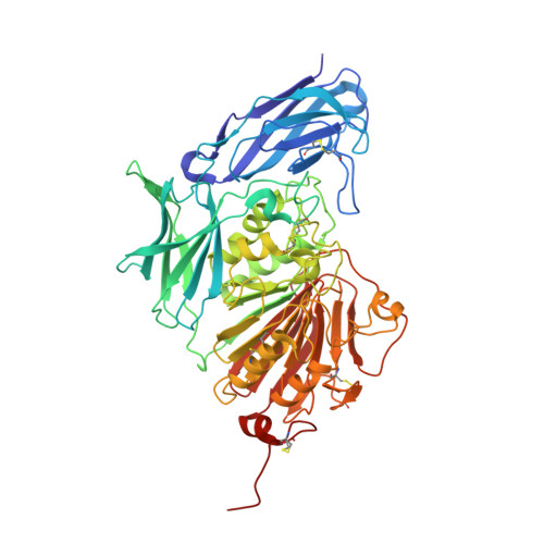
    Phosphatases function in the production, transport and recycling of inorganic phosphorus, which is crucial for cellular metabolism and bioenergetics, as well as in bacterial killing, since they are able to generate reactive oxygen species via Fenton chemistry. Diphosphonucleotide phosphatase/phosphodiesterase (PPD1), a glycoprotein plant purple acid phosphatase (PAP) from yellow lupin seeds, contains a bimetallic Fe-Mn catalytic site which is most active at acidic pH. Unlike other plant PAPs, PPD1 cleaves the pyrophosphate bond in diphosphonucleotides and the phosphodiester bond in various phosphodiesters. The homohexameric organization of PPD1, as revealed by a 1.65 Å resolution crystal structure and confirmed by solution X-ray scattering, is unique among plant PAPs, for which only homodimers have previously been reported. A phosphate anion is bound in a bidentate fashion at the active site, bridging the Fe and Mn atoms in a binding mode similar to that previously reported for sweet potato PAP, which suggests that common features occur in their catalytic mechanisms. The N-terminal domain of PPD1 has an unexpected and unique fibronectin type III-like fold that is absent in other plant PAPs. Here, the in vitro DNA-cleavage activity of PPD1 is demonstrated and it is proposed that the fibronectin III-like domain, which 'overhangs' the active site, is involved in DNA selectivity, binding and activation. The degradation of DNA by PPD1 implies a role for PPD1 in plant growth and repair and in pathogen defence.


  • Organizational Affiliation
    • Molecular Biophysics Group, Faculty of Health and Life Sciences, University of Liverpool , Crown Street, Liverpool L69 7ZB, England.

Macromolecule Content 

  • Total Structure Weight: 199.88 kDa 
  • Atom Count: 17,881 
  • Modeled Residue Count: 1,708 
  • Deposited Residue Count: 1,713 
  • Unique protein chains: 1

Macromolecules

Find similar proteins by:|  3D Structure
Entity ID: 1
MoleculeChains  Sequence LengthOrganismDetailsImage
DIPHOSPHONUCLEOTIDE PHOSPHATASE 1
A, B, C
571Lupinus luteusMutation(s): 0 
EC: 3.1.3.2
UniProt
Find proteins for Q8VX11 (Lupinus luteus)
Explore Q8VX11 
Go to UniProtKB:  Q8VX11
Entity Groups
Sequence Clusters30% Identity50% Identity70% Identity90% Identity95% Identity100% Identity
UniProt GroupQ8VX11
Glycosylation
Glycosylation Sites: 3
Sequence Annotations
Expand
Reference Sequence

Oligosaccharides

Help  
Entity ID: 2
MoleculeChains Length2D Diagram GlycosylationD Interactions
2-acetamido-2-deoxy-beta-D-glucopyranose-(1-4)-2-acetamido-2-deoxy-beta-D-glucopyranose
D, E, F, G, H
D, E, F, G, H, J
2N-Glycosylation
Glycosylation Resources
GlyTouCan: G42666HT
GlyCosmos: G42666HT
GlyGen: G42666HT
Entity ID: 3
MoleculeChains Length2D Diagram GlycosylationD Interactions
alpha-L-fucopyranose-(1-3)-[2-acetamido-2-deoxy-beta-D-glucopyranose-(1-4)]2-acetamido-2-deoxy-beta-D-glucopyranose
I, K
3N-Glycosylation
Glycosylation Resources
GlyTouCan: G11971MR
GlyCosmos: G11971MR
GlyGen: G11971MR

Small Molecules

Ligands 5 Unique
IDChains Name / Formula / InChI Key2D Diagram3D Interactions
NAG

Query on NAG


Download:Ideal Coordinates CCD File
AA [auth C]
BA [auth C]
CA [auth C]
O [auth A]
P [auth A]
AA [auth C],
BA [auth C],
CA [auth C],
O [auth A],
P [auth A],
V [auth B],
W [auth B]
2-acetamido-2-deoxy-beta-D-glucopyranose
C8 H15 N O6
OVRNDRQMDRJTHS-FMDGEEDCSA-N
PO4

Query on PO4


Download:Ideal Coordinates CCD File
DA [auth C],
Q [auth A],
X [auth B]
PHOSPHATE ION
O4 P
NBIIXXVUZAFLBC-UHFFFAOYSA-K
GOL

Query on GOL


Download:Ideal Coordinates CCD File
EA [auth C],
N [auth A],
R [auth A],
S [auth A]
GLYCEROL
C3 H8 O3
PEDCQBHIVMGVHV-UHFFFAOYSA-N
FE

Query on FE


Download:Ideal Coordinates CCD File
L [auth A],
T [auth B],
Y [auth C]
FE (III) ION
Fe
VTLYFUHAOXGGBS-UHFFFAOYSA-N
MN

Query on MN


Download:Ideal Coordinates CCD File
M [auth A],
U [auth B],
Z [auth C]
MANGANESE (II) ION
Mn
WAEMQWOKJMHJLA-UHFFFAOYSA-N

Experimental Data & Validation

Experimental Data

  • Method: X-RAY DIFFRACTION
  • Resolution: 1.65 Å
  • R-Value Free:  0.175 (Depositor), 0.183 (DCC) 
  • R-Value Work:  0.150 (Depositor), 0.161 (DCC) 
  • R-Value Observed: 0.151 (Depositor) 
Space Group: P 42 21 2
Unit Cell:
Length ( Å )Angle ( ˚ )
a = 223.925α = 90
b = 223.925β = 90
c = 111.904γ = 90
Software Package:
Software NamePurpose
REFMACrefinement
HKL-2000data reduction
HKL-2000data scaling
PHENIXphasing

Structure Validation

View Full Validation Report


Ligand Structure Quality Assessment 


Entry History 

Deposition Data

Revision History  (Full details and data files)

  • Version 1.0: 2014-01-29
    Type: Initial release
  • Version 1.1: 2014-03-12
    Changes: Database references, Other, Structure summary
  • Version 1.2: 2014-08-20
    Changes: Database references
  • Version 2.0: 2020-07-29
    Type: Remediation
    Reason: Carbohydrate remediation
    Changes: Advisory, Atomic model, Data collection, Derived calculations, Other, Structure summary
  • Version 2.1: 2024-10-09
    Changes: Data collection, Database references, Structure summary