3X0D | pdb_00003x0d

Crystal structure of C.elegans PRMT7 in complex with SAH (P43212)


Experimental Data Snapshot

  • Method: X-RAY DIFFRACTION
  • Resolution: 2.15 Å
  • R-Value Free: 
    0.266 (Depositor), 0.260 (DCC) 
  • R-Value Work: 
    0.220 (Depositor), 0.220 (DCC) 
  • R-Value Observed: 
    0.222 (Depositor) 

wwPDB Validation   3D Report Full Report


Ligand Structure Quality Assessment 

Created with Raphaël 2.3.0Worse 01 BetterLigand structure goodness of fit to experimental dataBest fitted SAHClick on this verticalbar to view details

This is version 1.1 of the entry. See complete history


Literature

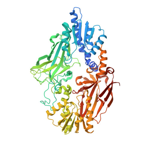
Protein arginine methyltransferase 7 has a novel homodimer-like structure formed by tandem repeats

Hasegawa, M.Toma-fukai, S.Kim, J.D.Fukamizu, A.Shimizu, T.

(2014) FEBS Lett 588: 1942-1948

  • DOI: https://doi.org/10.1016/j.febslet.2014.03.053
  • Primary Citation of Related Structures:  
    3WST, 3X0D

  • PubMed Abstract: 

    Protein arginine methyltransferase 7 (PRMT7) is a member of a family of enzymes that catalyze the transfer of methyl groups from S-adenosyl-l-methionine to nitrogen atoms on arginine residues. Here, we describe the crystal structure of Caenorhabditis elegans PRMT7 in complex with its reaction product S-adenosyl-L-homocysteine. The structural data indicated that PRMT7 harbors two tandem repeated PRMT core domains that form a novel homodimer-like structure. S-adenosyl-L-homocysteine bound to the N-terminal catalytic site only; the C-terminal catalytic site is occupied by a loop that inhibits cofactor binding. Mutagenesis demonstrated that only the N-terminal catalytic site of PRMT7 is responsible for cofactor binding.


  • Organizational Affiliation

    Graduate School of Pharmaceutical Sciences, The University of Tokyo, Hongo, Bunkyo-ku, Tokyo 113-0033, Japan.


Macromolecules
Find similar proteins by:  (by identity cutoff)  |  3D Structure
Entity ID: 1
MoleculeChains Sequence LengthOrganismDetailsImage
Protein arginine N-methyltransferase 7655Caenorhabditis elegansMutation(s): 0 
Gene Names: prmt-7pmrt-2W06D4.4
EC: 2.1.1
UniProt
Find proteins for Q9XW42 (Caenorhabditis elegans)
Explore Q9XW42 
Go to UniProtKB:  Q9XW42
Entity Groups  
Sequence Clusters30% Identity50% Identity70% Identity90% Identity95% Identity100% Identity
UniProt GroupQ9XW42
Sequence Annotations
Expand
  • Reference Sequence
Experimental Data & Validation

Experimental Data

  • Method: X-RAY DIFFRACTION
  • Resolution: 2.15 Å
  • R-Value Free:  0.266 (Depositor), 0.260 (DCC) 
  • R-Value Work:  0.220 (Depositor), 0.220 (DCC) 
  • R-Value Observed: 0.222 (Depositor) 
Space Group: P 43 21 2
Unit Cell:
Length ( Å )Angle ( ˚ )
a = 114.179α = 90
b = 114.179β = 90
c = 184.012γ = 90
Software Package:
Software NamePurpose
MOLREPphasing
REFMACrefinement
HKL-2000data reduction
HKL-2000data scaling

Structure Validation

View Full Validation Report


Ligand Structure Quality Assessment 

Created with Raphaël 2.3.0Worse 01 BetterLigand structure goodness of fit to experimental dataBest fitted SAHClick on this verticalbar to view details

Entry History 

Deposition Data

Revision History  (Full details and data files)

  • Version 1.0: 2014-10-29
    Type: Initial release
  • Version 1.1: 2024-03-20
    Changes: Data collection, Database references, Derived calculations