


Crystal structure of Pyrococcus horikoshii kynurenine aminotransferase in complex with PMP, GLA, 4AD, 2OG, GLU and KYA
Okada, K., Angkawidjaja, C., Koga, Y., Kanaya, S.To be published.
Experimental Data Snapshot
Starting Model: experimental
View more details
Entity ID: 1 | |||||
|---|---|---|---|---|---|
| Molecule | Chains | Sequence Length | Organism | Details | Image |
| Putative uncharacterized protein PH0207 | A, B [auth C] | 448 | Pyrococcus horikoshii OT3 | Mutation(s): 0 Gene Names: KAT, PH0207 | 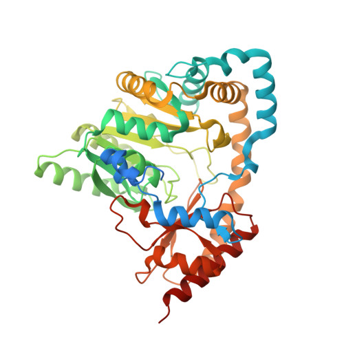 |
UniProt | |||||
Entity Groups | |||||
| Sequence Clusters | 30% Identity50% Identity70% Identity90% Identity95% Identity100% Identity | ||||
| UniProt Group | O57946 | ||||
Sequence AnnotationsExpand | |||||
Reference Sequence | |||||
| Ligands 6 Unique | |||||
|---|---|---|---|---|---|
| ID | Chains | Name / Formula / InChI Key | 2D Diagram | 3D Interactions | |
| PMP Download:Ideal Coordinates CCD File | C [auth A], H [auth C] | 4'-DEOXY-4'-AMINOPYRIDOXAL-5'-PHOSPHATE C8 H13 N2 O5 P ZMJGSOSNSPKHNH-UHFFFAOYSA-N |  | ||
| 3EE Download:Ideal Coordinates CCD File | F [auth A], K [auth C] | 4-(2-aminophenyl)-2,4-dioxobutanoic acid C10 H9 N O4 CAOVWYZQMPNAFJ-UHFFFAOYSA-N |  | ||
| KYA Download:Ideal Coordinates CCD File | L [auth C] | 4-hydroxyquinoline-2-carboxylic acid C10 H7 N O3 HCZHHEIFKROPDY-UHFFFAOYSA-N |  | ||
| GLU Download:Ideal Coordinates CCD File | G [auth A] | GLUTAMIC ACID C5 H9 N O4 WHUUTDBJXJRKMK-VKHMYHEASA-N |  | ||
| AKG Download:Ideal Coordinates CCD File | D [auth A], I [auth C] | 2-OXOGLUTARIC ACID C5 H6 O5 KPGXRSRHYNQIFN-UHFFFAOYSA-N |  | ||
| G9A Download:Ideal Coordinates CCD File | E [auth A], J [auth C] | (2E)-pent-2-enedioic acid C5 H6 O4 XVOUMQNXTGKGMA-OWOJBTEDSA-N |  | ||
| Length ( Å ) | Angle ( ˚ ) |
|---|---|
| a = 85.769 | α = 90 |
| b = 70.997 | β = 90.02 |
| c = 136.704 | γ = 90 |
| Software Name | Purpose |
|---|---|
| BL38B1 | data collection |
| MOLREP | phasing |
| REFMAC | refinement |
| HKL-2000 | data reduction |
| HKL-2000 | data scaling |


