3WMR

Crystal structure of VinJ


Experimental Data Snapshot

  • Method: X-RAY DIFFRACTION
  • Resolution: 1.95 Å
  • R-Value Free: 0.206 
  • R-Value Work: 0.165 
  • R-Value Observed: 0.167 

wwPDB Validation   3D Report Full Report


Ligand Structure Quality Assessment 


This is version 1.1 of the entry. See complete history


Literature

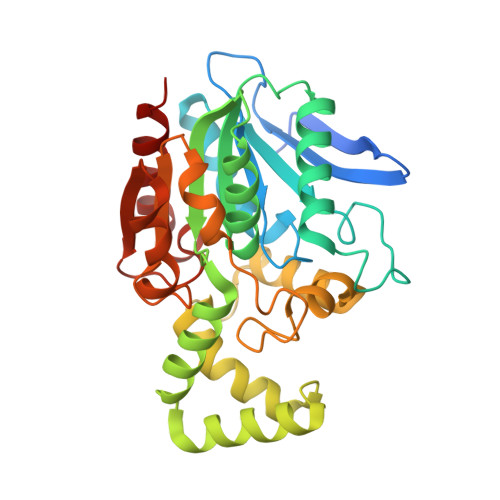
The crystal structure of the amidohydrolase VinJ shows a unique hydrophobic tunnel for its interaction with polyketide substrates

Shinohara, Y.Miyanaga, A.Kudo, F.Eguchi, T.

(2014) FEBS Lett 588: 995-1000

  • DOI: https://doi.org/10.1016/j.febslet.2014.01.060
  • Primary Citation of Related Structures:  
    3WMR

  • PubMed Abstract: 
  • VinJ is an amidohydrolase belonging to the serine peptidase family that catalyzes the hydrolysis of the terminal aminoacyl moiety of a polyketide intermediate during the biosynthesis of vicenistatin. Herein, we report the crystal structure of VinJ. VinJ possesses a unique hydrophobic tunnel for the recognition of the polyketide chain moiety of its substrate in the cap domain ...

    VinJ is an amidohydrolase belonging to the serine peptidase family that catalyzes the hydrolysis of the terminal aminoacyl moiety of a polyketide intermediate during the biosynthesis of vicenistatin. Herein, we report the crystal structure of VinJ. VinJ possesses a unique hydrophobic tunnel for the recognition of the polyketide chain moiety of its substrate in the cap domain. Taken together with the results of phylogenetic analysis, our results suggest that VinJ represents a new amidohydrolase family that is different from the known α/β hydrolase type serine peptidases.


    Organizational Affiliation

    Department of Chemistry and Materials Science, Tokyo Institute of Technology, O-okayama, Meguro-ku, Tokyo 152-8551, Japan. Electronic address: eguchi@cms.titech.ac.jp.


Macromolecules
Find similar proteins by:  (by identity cutoff)  |  3D Structure
Entity ID: 1
MoleculeChainsSequence LengthOrganismDetailsImage
Proline iminopeptidase
A, B, C
315Streptomyces halstediiMutation(s): 0 
Gene Names: vinJ
UniProt
Find proteins for Q76KY6 (Streptomyces halstedii)
Explore Q76KY6 
Go to UniProtKB:  Q76KY6
Entity Groups  
Sequence Clusters30% Identity50% Identity70% Identity90% Identity95% Identity100% Identity
UniProt GroupQ76KY6
Protein Feature View
Expand
  • Reference Sequence
Small Molecules
Ligands 2 Unique
IDChainsName / Formula / InChI Key2D Diagram3D Interactions
P4G
Query on P4G

Download Ideal Coordinates CCD File 
E [auth A],
H [auth B]
1-ETHOXY-2-(2-ETHOXYETHOXY)ETHANE
C8 H18 O3
RRQYJINTUHWNHW-UHFFFAOYSA-N
 Ligand Interaction
GOL
Query on GOL

Download Ideal Coordinates CCD File 
D [auth A],
F [auth B],
G [auth B]
GLYCEROL
C3 H8 O3
PEDCQBHIVMGVHV-UHFFFAOYSA-N
 Ligand Interaction
Experimental Data & Validation

Experimental Data

  • Method: X-RAY DIFFRACTION
  • Resolution: 1.95 Å
  • R-Value Free: 0.206 
  • R-Value Work: 0.165 
  • R-Value Observed: 0.167 
  • Space Group: P 1
Unit Cell:
Length ( Å )Angle ( ˚ )
a = 52.87α = 114.02
b = 72.62β = 98.39
c = 75.45γ = 98.5
Software Package:
Software NamePurpose
ADSCdata collection
MOLREPphasing
REFMACrefinement
MOSFLMdata reduction
SCALAdata scaling

Structure Validation

View Full Validation Report


Ligand Structure Quality Assessment 


Entry History 

Deposition Data

Revision History  (Full details and data files)

  • Version 1.0: 2014-02-05
    Type: Initial release
  • Version 1.1: 2014-03-19
    Changes: Database references