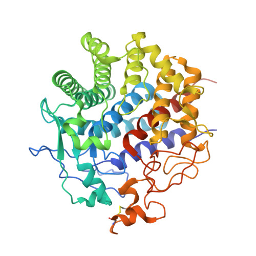
3WC3

Crystal structure of endo-1,4-beta-glucanase from Eisenia fetida


Experimental Data Snapshot

  • Method: X-RAY DIFFRACTION
  • Resolution: 1.50 Å
  • R-Value Free: 0.168 
  • R-Value Work: 0.147 
  • R-Value Observed: 0.148 

wwPDB Validation   3D Report Full Report


This is version 1.1 of the entry. See complete history


Literature

Crystal structure of endo-1,4-beta-glucanase from Eisenia fetida

Arimori, T.Ito, A.Nakazawa, M.Ueda, M.Tamada, T.

(2013) J Synchrotron Radiat 20: 884-889

  • DOI: https://doi.org/10.1107/S0909049513021110
  • Primary Citation of Related Structures:  
    3WC3

  • PubMed Abstract: 
  • The saccharification process is essential for bioethanol production from woody biomass including celluloses. Cold-adapted cellulase, which has sufficient activity at low temperature (<293 K), is capable of reducing heating costs during the saccharification process and is suitable for simultaneous saccharification and fermentation ...

    The saccharification process is essential for bioethanol production from woody biomass including celluloses. Cold-adapted cellulase, which has sufficient activity at low temperature (<293 K), is capable of reducing heating costs during the saccharification process and is suitable for simultaneous saccharification and fermentation. Endo-1,4-β-glucanase from the earthworm Eisenia fetida (EF-EG2) belonging to glycoside hydrolase family 9 has been shown to have the highest activity at 313 K, and also retained a comparatively high activity at 283 K. The recombinant EF-EG2 was purified expressed in Pichia pastoris, and then grew needle-shaped crystals with dimensions of 0.02 × 0.02 × 1 mm. The crystals belonged to the space group P3221 with unit-cell parameters of a = b = 136 Å, c = 55.0 Å. The final model of EF-EG2, including 435 residues, two ions, seven crystallization reagents and 696 waters, was refined to a crystallographic R-factor of 14.7% (free R-factor of 16.8%) to 1.5 Å resolution. The overall structure of EF-EG2 has an (α/α)6 barrel fold which contains a putative active-site cleft and a negatively charged surface. This structural information helps us understand the catalytic and cold adaptation mechanisms of EF-EG2.


    Organizational Affiliation

    Quantum Beam Science Directorate, Japan Atomic Energy Agency, 2-4 Shirakata-Shirane, Tokai, Ibaraki 319-1195, Japan.


Macromolecules
Find similar proteins by:  (by identity cutoff)  |  3D Structure
Entity ID: 1
MoleculeChainsSequence LengthOrganismDetailsImage
Endo-1, 4-beta-glucanase460Eisenia fetidaMutation(s): 0 
Gene Names: EF-EG2
EC: 3.2.1.4
Entity Groups  
Sequence Clusters30% Identity50% Identity70% Identity90% Identity95% Identity100% Identity
Protein Feature View
Expand
  • Reference Sequence
Small Molecules
Experimental Data & Validation

Experimental Data

  • Method: X-RAY DIFFRACTION
  • Resolution: 1.50 Å
  • R-Value Free: 0.168 
  • R-Value Work: 0.147 
  • R-Value Observed: 0.148 
  • Space Group: P 32 2 1
Unit Cell:
Length ( Å )Angle ( ˚ )
a = 136.449α = 90
b = 136.449β = 90
c = 54.974γ = 120
Software Package:
Software NamePurpose
SERGUIdata collection
PHASERphasing
REFMACrefinement
HKL-2000data reduction
HKL-2000data scaling

Structure Validation

View Full Validation Report


Entry History 

Deposition Data

Revision History  (Full details and data files)

  • Version 1.0: 2013-10-30
    Type: Initial release
  • Version 1.1: 2017-11-22
    Changes: Refinement description