3UJM

Crystal structure of the NTF2-like domain of the Drosophila melanogaster Rasputin protein


Experimental Data Snapshot

  • Method: X-RAY DIFFRACTION
  • Resolution: 2.74 Å
  • R-Value Free: 0.242 
  • R-Value Work: 0.197 
  • R-Value Observed: 0.199 

wwPDB Validation   3D Report Full Report


This is version 1.2 of the entry. See complete history


Literature

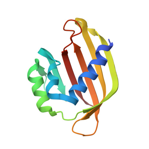
Crystal structure of the Rasputin NTF2-like domain from Drosophila melanogaster.

Vognsen, T.Kristensen, O.

(2012) Biochem Biophys Res Commun 420: 188-192

  • DOI: https://doi.org/10.1016/j.bbrc.2012.02.140
  • Primary Citation of Related Structures:  
    3UJM

  • PubMed Abstract: 

    The crystal structure of the NTF2-like domain of the Drosophila homolog of Ras GTPase SH3 Binding Protein (G3BP), Rasputin, was determined at 2.7Å resolution. The overall structure is highly similar to nuclear transport factor 2: It is a homodimer comprised of a β-sheet and three α-helices forming a cone-like shape. However, known binding sites for RanGDP and FxFG containing peptides show electrostatic and steric differences compared to nuclear transport factor 2. A HEPES molecule bound in the structure suggests a new, and possibly physiologically relevant, ligand binding site.


  • Organizational Affiliation

    Biostructural Research, Department of Drug Design and Pharmacology, Faculty of Health Sciences, University of Copenhagen, Universitetsparken 2, DK-2100 Copenhagen, Denmark. tv@farma.ku.dk


Macromolecules
Find similar proteins by:  (by identity cutoff)  |  3D Structure
Entity ID: 1
MoleculeChains Sequence LengthOrganismDetailsImage
Rasputin
A, B
120Drosophila melanogasterMutation(s): 0 
Gene Names: rinCG9412
UniProt
Find proteins for Q9VFT4 (Drosophila melanogaster)
Explore Q9VFT4 
Go to UniProtKB:  Q9VFT4
Entity Groups  
Sequence Clusters30% Identity50% Identity70% Identity90% Identity95% Identity100% Identity
UniProt GroupQ9VFT4
Sequence Annotations
Expand
  • Reference Sequence
Small Molecules
Ligands 1 Unique
IDChains Name / Formula / InChI Key2D Diagram3D Interactions
EPE
Query on EPE

Download Ideal Coordinates CCD File 
C [auth A],
D [auth B]
4-(2-HYDROXYETHYL)-1-PIPERAZINE ETHANESULFONIC ACID
C8 H18 N2 O4 S
JKMHFZQWWAIEOD-UHFFFAOYSA-N
Experimental Data & Validation

Experimental Data

  • Method: X-RAY DIFFRACTION
  • Resolution: 2.74 Å
  • R-Value Free: 0.242 
  • R-Value Work: 0.197 
  • R-Value Observed: 0.199 
  • Space Group: P 1 21 1
Unit Cell:
Length ( Å )Angle ( ˚ )
a = 39.2α = 90
b = 80.38β = 92.6
c = 39.2γ = 90
Software Package:
Software NamePurpose
xia2data scaling
XDSdata scaling
PHASERphasing
PHENIXrefinement
xia2data reduction
XDSdata reduction

Structure Validation

View Full Validation Report


Entry History 

Deposition Data

Revision History  (Full details and data files)

  • Version 1.0: 2012-04-25
    Type: Initial release
  • Version 1.1: 2017-11-08
    Changes: Refinement description
  • Version 1.2: 2023-09-13
    Changes: Data collection, Database references, Derived calculations, Refinement description