Structural and enzymatic analyses reveal the binding mode of a novel series of Francisella tularensis enoyl reductase (FabI) inhibitors.
Mehboob, S., Hevener, K.E., Truong, K., Boci, T., Santarsiero, B.D., Johnson, M.E.(2012) J Med Chem 55: 5933-5941
- PubMed: 22642319 Search on PubMedSearch on PubMed Central
- DOI: https://doi.org/10.1021/jm300489v
- Primary Citation Related Structures:
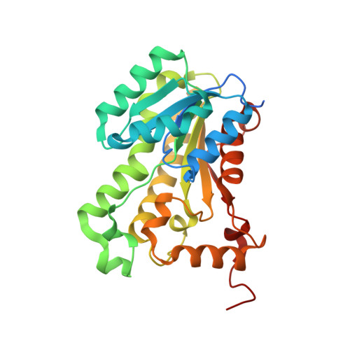
3UIC - PubMed Abstract:
Because of structural and mechanistic differences between eukaryotic and prokaryotic fatty acid synthesis enzymes, the bacterial pathway, FAS-II, is an attractive target for the design of antimicrobial agents. We have previously reported the identification of a novel series of benzimidazole compounds with particularly good antibacterial effect against Francisella tularensis, a Category A biowarfare pathogen. Herein we report the crystal structure of the F. tularensis FabI enzyme in complex with our most active benzimidazole compound bound with NADH. The structure reveals that the benzimidazole compounds bind to the substrate site in a unique conformation that is distinct from the binding motif of other known FabI inhibitors. Detailed inhibition kinetics have confirmed that the compounds possess a novel inhibitory mechanism that is unique among known FabI inhibitors. These studies could have a strong impact on future antimicrobial design efforts and may reveal new avenues for the design of FAS-II active antibacterial compounds.
- Center for Pharmaceutical Biotechnology, University of Illinois at Chicago, 900 S. Ashland Avenue, Chicago, Illinois 60607-7173, United States. mjohnson@uic.edu
Organizational Affiliation: