3UB5

Profilin:actin with a wide open nucleotide cleft


Experimental Data Snapshot

  • Method: X-RAY DIFFRACTION
  • Resolution: 2.20 Å
  • R-Value Free: 0.290 
  • R-Value Work: 0.215 
  • R-Value Observed: 0.219 

wwPDB Validation   3D Report Full Report


Ligand Structure Quality Assessment 


This is version 1.0 of the entry. See complete history


Literature

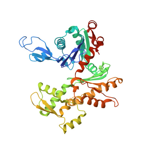
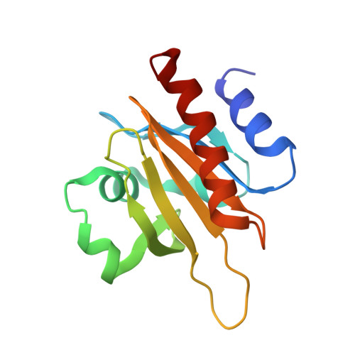
Structural basis for profilin-mediated actin nucleotide exchange.

Porta, J.C.Borgstahl, G.E.

(2012) J Mol Biol 418: 103-116

  • DOI: https://doi.org/10.1016/j.jmb.2012.02.012
  • Primary Citation of Related Structures:  
    3U4L, 3UB5

  • PubMed Abstract: 
  • Actin is a ubiquitous eukaryotic protein that is responsible for cellular scaffolding, motility, and division. The ability of actin to form a helical filament is the driving force behind these cellular activities. Formation of a filament depends on the successful exchange of actin's ADP for ATP ...

    Actin is a ubiquitous eukaryotic protein that is responsible for cellular scaffolding, motility, and division. The ability of actin to form a helical filament is the driving force behind these cellular activities. Formation of a filament depends on the successful exchange of actin's ADP for ATP. Mammalian profilin is a small actin binding protein that catalyzes the exchange of nucleotide and facilitates the addition of an actin monomer to a growing filament. Here, crystal structures of profilin-actin have been determined to show an actively exchanging ATP. Structural analysis shows how the binding of profilin to the barbed end of actin causes a rotation of the small domain relative to the large domain. This conformational change is propagated to the ATP site and causes a shift in nucleotide loops, which in turn causes a repositioning of Ca(2+) to its canonical position as the cleft closes around ATP. Reversal of the solvent exposure of Trp356 is also involved in cleft closure. In addition, secondary calcium binding sites were identified.


    Organizational Affiliation

    Department of Biochemistry and Molecular Biology, 987696 Nebraska Medical Center, Omaha, NE 68198-7696, USA.


Macromolecules
Find similar proteins by:  (by identity cutoff)  |  3D Structure
Entity ID: 1
MoleculeChainsSequence LengthOrganismDetailsImage
Actin, cytoplasmic 1374Bos taurusMutation(s): 0 
Gene Names: ACTB
UniProt
Find proteins for P60712 (Bos taurus)
Explore P60712 
Go to UniProtKB:  P60712
Entity Groups  
Sequence Clusters30% Identity50% Identity70% Identity90% Identity95% Identity100% Identity
UniProt GroupP60712
Protein Feature View
Expand
  • Reference Sequence
Find similar proteins by:  (by identity cutoff)  |  3D Structure
Entity ID: 2
MoleculeChainsSequence LengthOrganismDetailsImage
Profilin-1B [auth P]140Bos taurusMutation(s): 1 
Gene Names: PFN1
UniProt
Find proteins for P02584 (Bos taurus)
Explore P02584 
Go to UniProtKB:  P02584
Entity Groups  
Sequence Clusters30% Identity50% Identity70% Identity90% Identity95% Identity100% Identity
UniProt GroupP02584
Protein Feature View
Expand
  • Reference Sequence
Small Molecules
Ligands 4 Unique
IDChainsName / Formula / InChI Key2D Diagram3D Interactions
ATP
Query on ATP

Download Ideal Coordinates CCD File 
C [auth A]ADENOSINE-5'-TRIPHOSPHATE
C10 H16 N5 O13 P3
ZKHQWZAMYRWXGA-KQYNXXCUSA-N
 Ligand Interaction
GOL
Query on GOL

Download Ideal Coordinates CCD File 
F [auth P]GLYCEROL
C3 H8 O3
PEDCQBHIVMGVHV-UHFFFAOYSA-N
 Ligand Interaction
CA
Query on CA

Download Ideal Coordinates CCD File 
D [auth A],
E [auth A]
CALCIUM ION
Ca
BHPQYMZQTOCNFJ-UHFFFAOYSA-N
 Ligand Interaction
CL
Query on CL

Download Ideal Coordinates CCD File 
G [auth P]CHLORIDE ION
Cl
VEXZGXHMUGYJMC-UHFFFAOYSA-M
 Ligand Interaction
Experimental Data & Validation

Experimental Data

  • Method: X-RAY DIFFRACTION
  • Resolution: 2.20 Å
  • R-Value Free: 0.290 
  • R-Value Work: 0.215 
  • R-Value Observed: 0.219 
  • Space Group: P 21 21 21
Unit Cell:
Length ( Å )Angle ( ˚ )
a = 38α = 90
b = 72β = 90
c = 186.8γ = 90
Software Package:
Software NamePurpose
CrystalCleardata collection
REFMACrefinement
EVAL15data reduction
SADABSdata scaling

Structure Validation

View Full Validation Report


Ligand Structure Quality Assessment 


Entry History 

Deposition Data

Revision History  (Full details and data files)

  • Version 1.0: 2012-04-25
    Type: Initial release