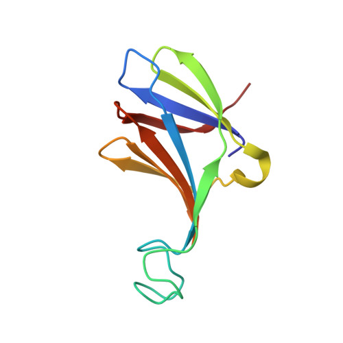
Structure of the phage TP901-1 1.8 MDa baseplate suggests an alternative host adhesion mechanism.
Veesler, D., Spinelli, S., Mahony, J., Lichiere, J., Blangy, S., Bricogne, G., Legrand, P., Ortiz-Lombardia, M., Campanacci, V., van Sinderen, D., Cambillau, C.(2012) Proc Natl Acad Sci U S A 109: 8954-8958
- PubMed: 22611190
- DOI: https://doi.org/10.1073/pnas.1200966109
- Primary Citation of Related Structures:
3U6X, 3UH8, 4V96 - PubMed Abstract:
Phages of the Caudovirales order possess a tail that recognizes the host and ensures genome delivery upon infection. The X-ray structure of the approximately 1.8 MDa host adsorption device (baseplate) from the lactococcal phage TP901-1 shows that the receptor-binding proteins are pointing in the direction of the host, suggesting that this organelle is in a conformation ready for host adhesion. This result is in marked contrast with the lactococcal phage p2 situation, whose baseplate is known to undergo huge conformational changes in the presence of Ca(2+) to reach its active state. In vivo infection experiments confirmed these structural observations by demonstrating that Ca(2+) ions are required for host adhesion among p2-like phages (936-species) but have no influence on TP901-1-like phages (P335-species). These data suggest that these two families rely on diverse adhesion strategies which may lead to different signaling for genome release.
- Architecture et Fonction des Macromolécules Biologiques, Aix-Marseille Université, Campus de Luminy, Case 932, 13288 Marseille Cedex 09, France. david.veesler@afmb.univ-mrs.fr
Organizational Affiliation:


