Pyrrolamide DNA gyrase inhibitors: fragment-based nuclear magnetic resonance screening to identify antibacterial agents.
Eakin, A.E., Green, O., Hales, N., Walkup, G.K., Bist, S., Singh, A., Mullen, G., Bryant, J., Embrey, K., Gao, N., Breeze, A., Timms, D., Andrews, B., Uria-Nickelsen, M., Demeritt, J., Loch, J.T., Hull, K., Blodgett, A., Illingworth, R.N., Prince, B., Boriack-Sjodin, P.A., Hauck, S., Macpherson, L.J., Ni, H., Sherer, B.(2012) Antimicrob Agents Chemother 56: 1240-1246
- PubMed: 22183167 Search on PubMedSearch on PubMed Central
- DOI: https://doi.org/10.1128/AAC.05485-11
- Primary Citation Related Structures:
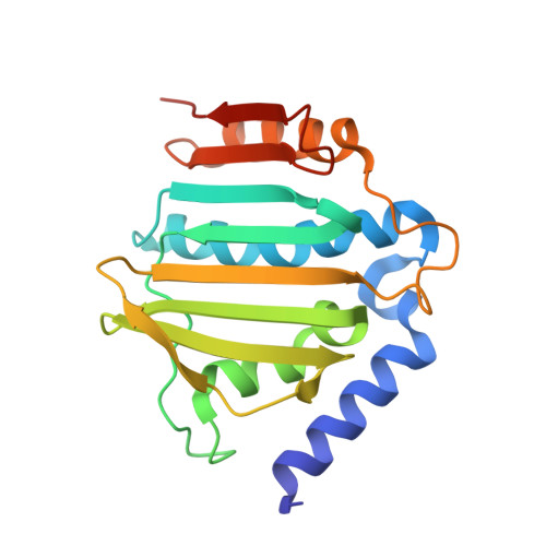
3U2D, 3U2K - PubMed Abstract:
DNA gyrase is an essential enzyme in bacteria, and its inhibition results in the disruption of DNA synthesis and, subsequently, cell death. The pyrrolamides are a novel class of antibacterial agents targeting DNA gyrase. These compounds were identified by a fragment-based lead generation (FBLG) approach using nuclear magnetic resonance (NMR) screening to identify low-molecular-weight compounds that bind to the ATP pocket of DNA gyrase. A pyrrole hit with a binding constant of 1 mM formed the basis of the design and synthesis of a focused library of compounds that resulted in the rapid identification of a lead compound that inhibited DNA gyrase with a 50% inhibitory concentration (IC(50)) of 3 μM. The potency of the lead compound was further optimized by utilizing iterative X-ray crystallography to yield DNA gyrase inhibitors that also displayed antibacterial activity. Spontaneous mutants were isolated in Staphylococcus aureus by plating on agar plates containing pyrrolamide 4 at the MIC. The resistant variants displayed 4- to 8-fold-increased MIC values relative to the parent strain. DNA sequencing revealed two independent point mutations in the pyrrolamide binding region of the gyrB genes from these variants, supporting the hypothesis that the mode of action of these compounds was inhibition of DNA gyrase. Efficacy of a representative pyrrolamide was demonstrated against Streptococcus pneumoniae in a mouse lung infection model. These data demonstrate that the pyrrolamides are a novel class of DNA gyrase inhibitors with the potential to deliver future antibacterial agents targeting multiple clinical indications.
- Infection Innovative Medicines Unit, AstraZeneca R&D Boston, Waltham, Massachusetts, USA. Ann.Eakin@astrazeneca.com
Organizational Affiliation: