3TXN

Crystal structure of Rpn6 from Drosophila melanogaster, native data


Experimental Data Snapshot

  • Method: X-RAY DIFFRACTION
  • Resolution: 2.50 Å
  • R-Value Free: 0.265 
  • R-Value Work: 0.216 
  • R-Value Observed: 0.218 

wwPDB Validation   3D Report Full Report


This is version 1.3 of the entry. See complete history


Literature

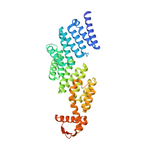
The proteasomal subunit Rpn6 is a molecular clamp holding the core and regulatory subcomplexes together.

Pathare, G.R.Nagy, I.Bohn, S.Unverdorben, P.Hubert, A.Korner, R.Nickell, S.Lasker, K.Sali, A.Tamura, T.Nishioka, T.Forster, F.Baumeister, W.Bracher, A.

(2012) Proc Natl Acad Sci U S A 109: 149-154

  • DOI: https://doi.org/10.1073/pnas.1117648108
  • Primary Citation of Related Structures:  
    3TXM, 3TXN

  • PubMed Abstract: 

    Proteasomes execute the degradation of most cellular proteins. Although the 20S core particle (CP) has been studied in great detail, the structure of the 19S regulatory particle (RP), which prepares ubiquitylated substrates for degradation, has remained elusive. Here, we report the crystal structure of one of the RP subunits, Rpn6, and we describe its integration into the cryo-EM density map of the 26S holocomplex at 9.1 Å resolution. Rpn6 consists of an α-solenoid-like fold and a proteasome COP9/signalosome eIF3 (PCI) module in a right-handed suprahelical configuration. Highly conserved surface areas of Rpn6 interact with the conserved surfaces of the Pre8 (alpha2) and Rpt6 subunits from the alpha and ATPase rings, respectively. The structure suggests that Rpn6 has a pivotal role in stabilizing the otherwise weak interaction between the CP and the RP.


  • Organizational Affiliation

    Department of Molecular Structural Biology, Max-Planck-Institute of Biochemistry, Am Klopferspitz 18, 82152 Martinsried, Germany.


Macromolecules
Find similar proteins by:  (by identity cutoff)  |  3D Structure
Entity ID: 1
MoleculeChains Sequence LengthOrganismDetailsImage
26S proteasome regulatory complex subunit p42B394Drosophila melanogasterMutation(s): 0 
Gene Names: BcDNA.LD18931CG10149Dmel_CG10149Rpn6
UniProt
Find proteins for Q7KLV9 (Drosophila melanogaster)
Explore Q7KLV9 
Go to UniProtKB:  Q7KLV9
Entity Groups  
Sequence Clusters30% Identity50% Identity70% Identity90% Identity95% Identity100% Identity
UniProt GroupQ7KLV9
Sequence Annotations
Expand
  • Reference Sequence
Experimental Data & Validation

Experimental Data

  • Method: X-RAY DIFFRACTION
  • Resolution: 2.50 Å
  • R-Value Free: 0.265 
  • R-Value Work: 0.216 
  • R-Value Observed: 0.218 
  • Space Group: P 61
Unit Cell:
Length ( Å )Angle ( ˚ )
a = 161.251α = 90
b = 161.251β = 90
c = 42.08γ = 120
Software Package:
Software NamePurpose
SCALAdata scaling
REFMACrefinement
PDB_EXTRACTdata extraction
XDSdata reduction
SHELXDphasing

Structure Validation

View Full Validation Report


Entry History 

Deposition Data

Revision History  (Full details and data files)

  • Version 1.0: 2011-12-14
    Type: Initial release
  • Version 1.1: 2012-01-11
    Changes: Database references
  • Version 1.2: 2012-02-01
    Changes: Database references
  • Version 1.3: 2024-02-28
    Changes: Data collection, Database references, Derived calculations