3TTY

Crystal structure of beta-galactosidase from Bacillus circulans sp. alkalophilus in complex with galactose


Experimental Data Snapshot

  • Method: X-RAY DIFFRACTION
  • Resolution: 2.25 Å
  • R-Value Free: 0.210 
  • R-Value Work: 0.160 
  • R-Value Observed: 0.163 

wwPDB Validation   3D Report Full Report


Ligand Structure Quality Assessment 


This is version 1.3 of the entry. See complete history


Literature

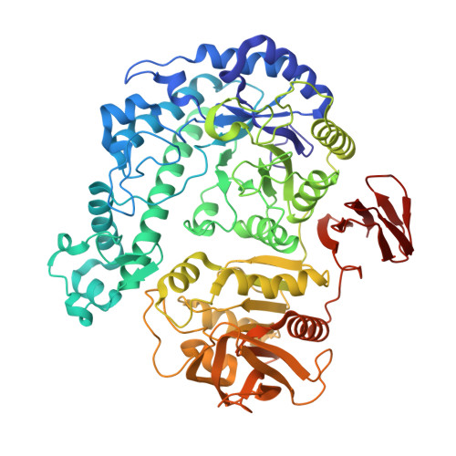
Structural analysis, enzymatic characterization, and catalytic mechanisms of beta-galactosidase from Bacillus circulans sp. alkalophilus.

Maksimainen, M.Paavilainen, S.Hakulinen, N.Rouvinen, J.

(2012) FEBS J 279: 1788-1798

  • DOI: https://doi.org/10.1111/j.1742-4658.2012.08555.x
  • Primary Citation of Related Structures:  
    3TTS, 3TTY

  • PubMed Abstract: 

    Crystal structures of native and α-D-galactose-bound Bacillus circulans sp. alkalophilus β-galactosidase (Bca-β-gal) were determined at 2.40 and 2.25 Å resolutions, respectively. Bca-β-gal is a member of family 42 of glycoside hydrolases, and forms a 460 kDa hexameric structure in crystal. The protein consists of three domains, of which the catalytic domain has an (α/β)(8) barrel structure with a cluster of sulfur-rich residues inside the β-barrel. The shape of the active site is clearly more open compared to the only homologous structure available in the Protein Data Bank. This is due to the number of large differences in the loops that connect the C-terminal ends of the β-strands to the N-terminal ends of the α-helices within the (α/β)(8) barrel. The complex structure shows that galactose binds to the active site as an α-anomer and induces clear conformational changes in the active site. The implications of α-D-galactose binding with respect to the catalytic mechanism are discussed. In addition, we suggest that β-galactosidases mainly utilize a reverse hydrolysis mechanism for synthesis of galacto-oligosaccharides.


  • Organizational Affiliation

    Department of Chemistry, University of Eastern Finland, Joensuu, Finland.


Macromolecules
Find similar proteins by:  (by identity cutoff)  |  3D Structure
Entity ID: 1
MoleculeChains Sequence LengthOrganismDetailsImage
Beta-galactosidase
A, B, C, D, E
A, B, C, D, E, F
675Niallia circulans subsp. alkalophilusMutation(s): 0 
EC: 3.2.1.23
Entity Groups  
Sequence Clusters30% Identity50% Identity70% Identity90% Identity95% Identity100% Identity
Sequence Annotations
Expand
  • Reference Sequence
Small Molecules
Ligands 2 Unique
IDChains Name / Formula / InChI Key2D Diagram3D Interactions
GLA
Query on GLA

Download Ideal Coordinates CCD File 
H [auth A]
J [auth B]
L [auth C]
N [auth D]
P [auth E]
H [auth A],
J [auth B],
L [auth C],
N [auth D],
P [auth E],
R [auth F]
alpha-D-galactopyranose
C6 H12 O6
WQZGKKKJIJFFOK-PHYPRBDBSA-N
ZN
Query on ZN

Download Ideal Coordinates CCD File 
G [auth A]
I [auth B]
K [auth C]
M [auth D]
O [auth E]
G [auth A],
I [auth B],
K [auth C],
M [auth D],
O [auth E],
Q [auth F]
ZINC ION
Zn
PTFCDOFLOPIGGS-UHFFFAOYSA-N
Experimental Data & Validation

Experimental Data

  • Method: X-RAY DIFFRACTION
  • Resolution: 2.25 Å
  • R-Value Free: 0.210 
  • R-Value Work: 0.160 
  • R-Value Observed: 0.163 
  • Space Group: H 3
Unit Cell:
Length ( Å )Angle ( ˚ )
a = 228.2α = 90
b = 228.2β = 90
c = 246.8γ = 120
Software Package:
Software NamePurpose
ADSCdata collection
PHASERphasing
PHENIXrefinement
XDSdata reduction
XSCALEdata scaling

Structure Validation

View Full Validation Report


Ligand Structure Quality Assessment 


Entry History 

Deposition Data

Revision History  (Full details and data files)

  • Version 1.0: 2012-03-21
    Type: Initial release
  • Version 1.1: 2013-06-19
    Changes: Database references
  • Version 1.2: 2020-07-29
    Type: Remediation
    Reason: Carbohydrate remediation
    Changes: Data collection, Derived calculations, Structure summary
  • Version 1.3: 2023-09-13
    Changes: Data collection, Database references, Refinement description, Structure summary