Crystal structure and immunologic characterization of the major grass pollen allergen Phl p 4.
Zafred, D., Nandy, A., Pump, L., Kahlert, H., Keller, W.(2013) J Allergy Clin Immunol 132: 696-703.e10
- PubMed: 23683465 Search on PubMed
- DOI: https://doi.org/10.1016/j.jaci.2013.03.021
- Primary Citation Related Structures:
3TSH, 3TSJ - PubMed Abstract:
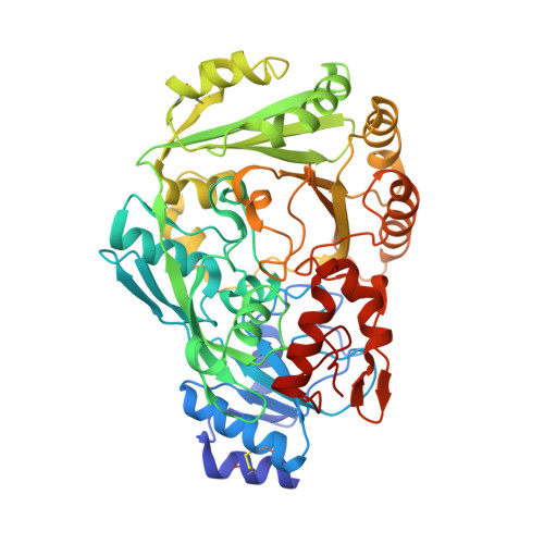
Phl p 4 is a major pollen allergen but exhibits lower allergenicity than other major allergens. The natural protein is glycosylated and shows cross-reactivity with related and structurally unrelated allergens. We sought to determine the high-resolution crystal structure of Phl p 4 and to evaluate the immunologic properties of the recombinant allergen in comparison with natural Phl p 4. Different isoallergens of Phl p 4 were expressed, and the nonglycosylated mutant was crystallized. The specific role of protein and carbohydrate epitopes for allergenicity was studied by using IgE inhibition and basophil release assays. The 3-dimensional structure was determined by using x-ray crystallography at a resolution of 1.9 Å. The allergen is a glucose dehydrogenase with a bicovalently attached flavin adenine dinucleotide. Glycosylated and nonglycosylated recombinant Phl p 4 showed identical inhibition of IgE binding, but compared with natural Phl p 4, all recombinant isoforms displayed a reduced IgE-binding inhibition. However, the recombinant protein exhibited an approximately 10-fold higher potency in basophil release assays than the natural protein. The crystal structure reveals the compact globular nature of the protein, and the observed binding pocket implies the size of the natural substrate. Plant-derived cross-reactive carbohydrate determinants (CCDs) appear to reduce the allergenicity of the natural allergen, whereas the Pichia pastoris-derived glycosylation does not. Our results imply yet undescribed mechanism of how CCDs dampen the immune response, leading to a novel understanding of the role of CCDs.
- Institute for Molecular Biosciences (IMB), University of Graz, Graz, Austria.
Organizational Affiliation: