3TJZ | pdb_00003tjz

Crystal Structure of Arf1 Bound to the gamma/zeta-COP Core Complex


Experimental Data Snapshot

  • Method: X-RAY DIFFRACTION
  • Resolution: 2.90 Å
  • R-Value Free: 
    0.260 (Depositor), 0.270 (DCC) 
  • R-Value Work: 
    0.206 (Depositor), 0.220 (DCC) 
  • R-Value Observed: 
    0.209 (Depositor) 

wwPDB Validation   3D Report Full Report


Ligand Structure Quality Assessment 

Created with Raphaël 2.3.0Worse 01 BetterLigand structure goodness of fit to experimental dataBest fitted GNPClick on this verticalbar to view details

This is version 1.1 of the entry. See complete history


Literature

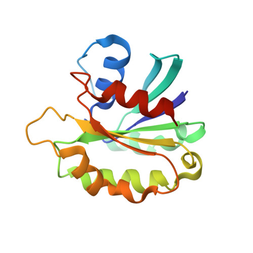
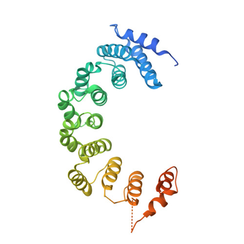
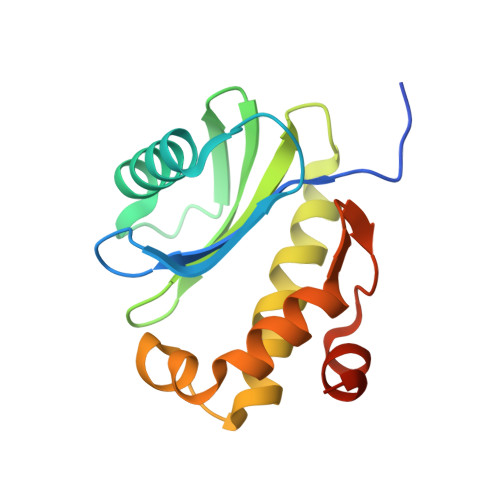
A structure-based mechanism for arf1-dependent recruitment of coatomer to membranes.

Yu, X.Breitman, M.Goldberg, J.

(2012) Cell 148: 530-542

  • DOI: https://doi.org/10.1016/j.cell.2012.01.015
  • Primary Citation of Related Structures:  
    3TJZ

  • PubMed Abstract: 

    Budding of COPI-coated vesicles from Golgi membranes requires an Arf family G protein and the coatomer complex recruited from cytosol. Arf is also required with coatomer-related clathrin adaptor complexes to bud vesicles from the trans-Golgi network and endosomal compartments. To understand the structural basis for Arf-dependent recruitment of a vesicular coat to the membrane, we determined the structure of Arf1 bound to the γζ-COP subcomplex of coatomer. Structure-guided biochemical analysis reveals that a second Arf1-GTP molecule binds to βδ-COP at a site common to the γ- and β-COP subunits. The Arf1-binding sites on coatomer are spatially related to PtdIns4,5P(2)-binding sites on the endocytic AP2 complex, providing evidence that the orientation of membrane binding is general for this class of vesicular coat proteins. A bivalent GTP-dependent binding mode has implications for the dynamics of coatomer interaction with the Golgi and for the selection of cargo molecules.


  • Organizational Affiliation

    Howard Hughes Medical Institute and the Structural Biology Program, Memorial Sloan-Kettering Cancer Center, 1275 York Avenue, New York, NY 10065, USA.


Macromolecules
Find similar proteins by:  (by identity cutoff)  |  3D Structure
Entity ID: 1
MoleculeChains Sequence LengthOrganismDetailsImage
ADP-ribosylation factor 1
A, D
164Saccharomyces cerevisiae S288CMutation(s): 0 
Gene Names: ARF1YDL192WD1244
EC: 3.6.5.2
UniProt
Find proteins for P11076 (Saccharomyces cerevisiae (strain ATCC 204508 / S288c))
Explore P11076 
Go to UniProtKB:  P11076
Entity Groups  
Sequence Clusters30% Identity50% Identity70% Identity90% Identity95% Identity100% Identity
UniProt GroupP11076
Sequence Annotations
Expand
  • Reference Sequence
Find similar proteins by:  (by identity cutoff)  |  3D Structure
Entity ID: 2
MoleculeChains Sequence LengthOrganismDetailsImage
Coatomer subunit gamma
B, E
355Bos taurusMutation(s): 0 
Gene Names: COPG
UniProt
Find proteins for P53620 (Bos taurus)
Explore P53620 
Go to UniProtKB:  P53620
Entity Groups  
Sequence Clusters30% Identity50% Identity70% Identity90% Identity95% Identity100% Identity
UniProt GroupP53620
Sequence Annotations
Expand
  • Reference Sequence
Find similar proteins by:  (by identity cutoff)  |  3D Structure
Entity ID: 3
MoleculeChains Sequence LengthOrganismDetailsImage
Coatomer subunit zeta-1
C, F
153Bos taurusMutation(s): 0 
Gene Names: COPZ1COPZ
UniProt
Find proteins for P35604 (Bos taurus)
Explore P35604 
Go to UniProtKB:  P35604
Entity Groups  
Sequence Clusters30% Identity50% Identity70% Identity90% Identity95% Identity100% Identity
UniProt GroupP35604
Sequence Annotations
Expand
  • Reference Sequence
Experimental Data & Validation

Experimental Data

  • Method: X-RAY DIFFRACTION
  • Resolution: 2.90 Å
  • R-Value Free:  0.260 (Depositor), 0.270 (DCC) 
  • R-Value Work:  0.206 (Depositor), 0.220 (DCC) 
  • R-Value Observed: 0.209 (Depositor) 
Space Group: P 42 21 2
Unit Cell:
Length ( Å )Angle ( ˚ )
a = 163.579α = 90
b = 163.579β = 90
c = 145.188γ = 90
Software Package:
Software NamePurpose
CBASSdata collection
PHENIXmodel building
PHENIXrefinement
HKL-2000data reduction
HKL-2000data scaling
PHENIXphasing

Structure Validation

View Full Validation Report


Ligand Structure Quality Assessment 

Created with Raphaël 2.3.0Worse 01 BetterLigand structure goodness of fit to experimental dataBest fitted GNPClick on this verticalbar to view details

Entry History 

Deposition Data

Revision History  (Full details and data files)

  • Version 1.0: 2012-02-22
    Type: Initial release
  • Version 1.1: 2024-02-28
    Changes: Data collection, Database references, Derived calculations