Binding of alpha , alpha-disubstituted amino acids to arginase suggests new avenues for inhibitor design.
Ilies, M., Di Costanzo, L., Dowling, D.P., Thorn, K.J., Christianson, D.W.(2011) J Med Chem 54: 5432-5443
- PubMed: 21728378 Search on PubMedSearch on PubMed Central
- DOI: https://doi.org/10.1021/jm200443b
- Primary Citation Related Structures:
3GMZ, 3GN0, 3SJT, 3SKK, 3SL0, 3SL1 - PubMed Abstract:
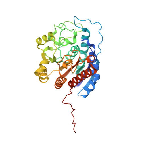
Arginase is a binuclear manganese metalloenzyme that hydrolyzes L-arginine to form L-ornithine and urea, and aberrant arginase activity is implicated in various diseases such as erectile dysfunction, asthma, atherosclerosis, and cerebral malaria. Accordingly, arginase inhibitors may be therapeutically useful. Continuing our efforts to expand the chemical space of arginase inhibitor design and inspired by the binding of 2-(difluoromethyl)-L-ornithine to human arginase I, we now report the first study of the binding of α,α-disubstituted amino acids to arginase. Specifically, we report the design, synthesis, and assay of racemic 2-amino-6-borono-2-methylhexanoic acid and racemic 2-amino-6-borono-2-(difluoromethyl)hexanoic acid. X-ray crystal structures of human arginase I and Plasmodium falciparum arginase complexed with these inhibitors reveal the exclusive binding of the L-stereoisomer; the additional α-substituent of each inhibitor is readily accommodated and makes new intermolecular interactions in the outer active site of each enzyme. Therefore, this work highlights a new region of the protein surface that can be targeted for additional affinity interactions, as well as the first comparative structural insights on inhibitor discrimination between a human and a parasitic arginase.
- Department of Chemistry, Drexel University, Philadelphia, Pennsylvania 19104-2875, United States.
Organizational Affiliation: