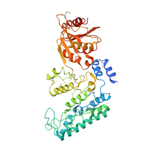
A Novel N-acetylglutamate synthase architecture revealed by the crystal structure of the bifunctional enzyme from Maricaulis maris.
Shi, D., Li, Y., Cabrera-Luque, J., Jin, Z., Yu, X., Zhao, G., Haskins, N., Allewell, N.M., Tuchman, M.(2011) PLoS One 6: e28825-e28825
- PubMed: 22174908 Search on PubMedSearch on PubMed Central
- DOI: https://doi.org/10.1371/journal.pone.0028825
- Primary Citation Related Structures:
3S6G, 3S6H, 3S6K, 3S7Y - PubMed Abstract:
Novel bifunctional N-acetylglutamate synthase/kinases (NAGS/K) that catalyze the first two steps of arginine biosynthesis and are homologous to vertebrate N-acetylglutamate synthase (NAGS), an essential cofactor-producing enzyme in the urea cycle, were identified in Maricaulis maris and several other bacteria. Arginine is an allosteric inhibitor of NAGS but not NAGK activity. The crystal structure of M. maris NAGS/K (mmNAGS/K) at 2.7 Å resolution indicates that it is a tetramer, in contrast to the hexameric structure of Neisseria gonorrhoeae NAGS. The quaternary structure of crystalline NAGS/K from Xanthomonas campestris (xcNAGS/K) is similar, and cross-linking experiments indicate that both mmNAGS/K and xcNAGS are tetramers in solution. Each subunit has an amino acid kinase (AAK) domain, which is likely responsible for N-acetylglutamate kinase (NAGK) activity and has a putative arginine binding site, and an N-acetyltransferase (NAT) domain that contains the putative NAGS active site. These structures and sequence comparisons suggest that the linker residue 291 may determine whether arginine acts as an allosteric inhibitor or activator in homologous enzymes in microorganisms and vertebrates. In addition, the angle of rotation between AAK and NAT domains varies among crystal forms and subunits within the tetramer. A rotation of 26° is sufficient to close the predicted AcCoA binding site, thus reducing enzymatic activity. Since mmNAGS/K has the highest degree of sequence homology to vertebrate NAGS of NAGS and NAGK enzymes whose structures have been determined, the mmNAGS/K structure was used to develop a structural model of human NAGS that is fully consistent with the functional effects of the 14 missense mutations that were identified in NAGS-deficient patients.
- Center for Genetic Medicine Research and Department of Integrative Systems Biology, Children's National Medical Center, The George Washington University, Washington, DC, United States of America. dshi@cnmcresearch.org
Organizational Affiliation: