


Crystal structure of putative amidohydrolase-2 (EFI-target 500288)from Polaromonas sp. JS666
Ramagopal, U.A., Toro, R., Girlt, J.A., Almo, S.C.To be published.
Experimental Data Snapshot
Starting Model: experimental
View more details
wwPDB Validation 3D Report Full Report

Macromolecule Content
Entity ID: 1 | |||||
|---|---|---|---|---|---|
| Molecule | Chains | Sequence Length | Organism | Details | Image |
| Amidohydrolase 2 | 348 | Polaromonas sp. JS666 | Mutation(s): 0 Gene Names: Bpro_2061 EC: 4.1.1.103 | 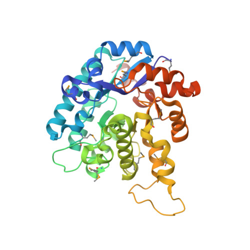 | |
UniProt | |||||
Entity Groups | |||||
| Sequence Clusters | 30% Identity50% Identity70% Identity90% Identity95% Identity100% Identity | ||||
| UniProt Group | Q12BV1 | ||||
Sequence AnnotationsExpand | |||||
Reference Sequence | |||||
| Ligands 5 Unique | |||||
|---|---|---|---|---|---|
| ID | Chains | Name / Formula / InChI Key | 2D Diagram | 3D Interactions | |
| EPE Download:Ideal Coordinates CCD File | DA [auth D] I [auth A] Q [auth B] QA [auth F] RA [auth F] | 4-(2-HYDROXYETHYL)-1-PIPERAZINE ETHANESULFONIC ACID C8 H18 N2 O4 S JKMHFZQWWAIEOD-UHFFFAOYSA-N |  | ||
| SO4 Download:Ideal Coordinates CCD File | AB [auth G] BB [auth G] CB [auth G] DB [auth G] EA [auth D] | SULFATE ION O4 S QAOWNCQODCNURD-UHFFFAOYSA-L |  | ||
| GOL Download:Ideal Coordinates CCD File | CA [auth C], JA [auth D], VA [auth F] | GLYCEROL C3 H8 O3 PEDCQBHIVMGVHV-UHFFFAOYSA-N |  | ||
| ACT Download:Ideal Coordinates CCD File | AA [auth C] BA [auth C] EB [auth G] FB [auth G] GB [auth G] | ACETATE ION C2 H3 O2 QTBSBXVTEAMEQO-UHFFFAOYSA-M |  | ||
| CL Download:Ideal Coordinates CCD File | X [auth B] | CHLORIDE ION Cl VEXZGXHMUGYJMC-UHFFFAOYSA-M |  | ||
| Modified Residues 1 Unique | |||||
|---|---|---|---|---|---|
| ID | Chains | Type | Formula | 2D Diagram | Parent |
| MSE Query on MSE | A, B, C, D, E A, B, C, D, E, F, G, H | L-PEPTIDE LINKING | C5 H11 N O2 Se |  | MET |
| Length ( Å ) | Angle ( ˚ ) |
|---|---|
| a = 81.024 | α = 90 |
| b = 151.241 | β = 91.96 |
| c = 143.41 | γ = 90 |
| Software Name | Purpose |
|---|---|
| SCALEPACK | data scaling |
| REFMAC | refinement |
| PDB_EXTRACT | data extraction |
| CBASS | data collection |
| HKL-2000 | data reduction |
| MOLREP | phasing |


