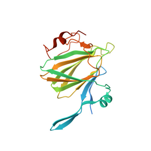
Structure of the Bacillus anthracis dTDP-L-rhamnose-biosynthetic enzyme dTDP-4-dehydrorhamnose 3,5-epimerase (RfbC).
Shornikov, A., Tran, H., Macias, J., Halavaty, A.S., Minasov, G., Anderson, W.F., Kuhn, M.L.(2017) Acta Crystallogr F Struct Biol Commun 73: 664-671
- PubMed: 29199987 Search on PubMedSearch on PubMed Central
- DOI: https://doi.org/10.1107/S2053230X17015849
- Primary Citation Related Structures:
3RYK - PubMed Abstract:
The exosporium layer of Bacillus anthracis spores is rich in L-rhamnose, a common bacterial cell-wall component, which often contributes to the virulence of pathogens by increasing their adherence and immune evasion. The biosynthetic pathway used to form the activated L-rhamnose donor dTDP-L-rhamnose consists of four enzymes (RfbA, RfbB, RfbC and RfbD) and is an attractive drug target because there are no homologs in mammals. It was found that co-purifying and screening RfbC (dTDP-6-deoxy-D-xylo-4-hexulose 3,5-epimerase) from B. anthracis in the presence of the other three B. anthracis enzymes of the biosynthetic pathway yielded crystals that were suitable for data collection. RfbC crystallized as a dimer and its structure was determined at 1.63 Å resolution. Two different ligands were bound in the protein structure: pyrophosphate in the active site of one monomer and dTDP in the other monomer. A structural comparison with RfbC homologs showed that the key active-site residues are conserved across kingdoms.
- Department of Chemistry and Biochemistry, San Francisco State University, USA.
Organizational Affiliation: